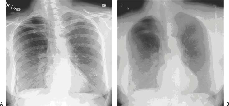
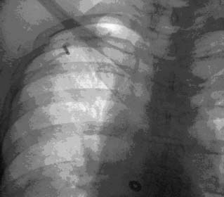
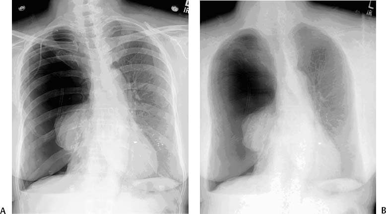
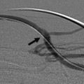
CASE 46 A 48-year-old female experienced shortness of breath 20 minutes after percutaneous chest biopsy. Figure 46-1 (A) Frontal chest radiograph following percutaneous biopsy shows a 5-cm apical pneumothorax. (B) Dual energy radiography minimizes the appearance of the overlying bony structures and emphasizes the pneumothorax and right apical 2-cm nodule. Chest radiograph showed a 5-cm apical pneumothorax of the right lung (Fig. 46–1). Pneumothorax complicating percutaneous chest biopsy. The patient received immediate supplemental oxygen by nasal cannula. Heart rate, respiratory rate, blood pressure, and oxygen saturation were monitored. She was placed supine on the fluoroscopy table. The anterolateral interspace between the first and second ribs was punctured under direct fluoroscopic guidance using the Trocar technique. A 6.3-French (F) pigtail catheter (Turner pneumothorax kit, Cook, Bloomington, Indiana) was advanced over a sharp stylet into the pleural space, and the pigtail catheter was advanced off the stylet. The pleural space was evacuated using a 60-cc syringe (Fig. 46-2), and the catheter was attached to a one-way Heimlich valve. The patient was admitted to the interventional radiology service for overnight observation. Four hours after admission, she reported increasing chest tightness, and radiography revealed tension pneumothorax of the right lung (Fig. 46-3). She was immediately recalled to interventional radiology and found to have an obstructed pleural drain. Patency of the drain was restored by manipulation under fluoroscopy (Fig. 46-4). The following morning, complete resolution of the pneumothorax was observed. The catheter was removed, and a radiograph obtained 2 hours after removal showed no evidence of recurrence. Figure 46-2 Fluoroscopic image shows placement of a 6F Turner pigtail catheter (Cook, Bloomington, Indiana) and re-expansion of the lung. Figure 46-3 (A) Frontal chest radiograph obtained after the patient complained of increasing chest tightness shows tension pneumothorax with complete collapse of the right lung. (B) Dual energy radiograph emphasizes the finding. Turner pneumothorax kit (Cook, Bloomington, Indiana) (Fig. 46-5)
Clinical Presentation
Radiologic Studies
Diagnosis
Treatment
Equipment
Discussion
Background
Related posts:


Stay updated, free articles. Join our Telegram channel

Full access? Get Clinical Tree


