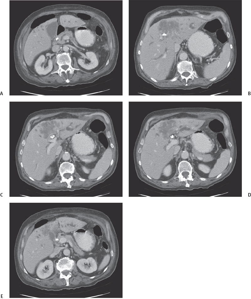
CASE 7 A 68-year-old man presents with jaundice, weight loss, and anorexia. Fig. 7.1 (A–E) Contrast-enhanced CT image of liver parenchyma reveals an extensive, ill-defined, hypodense mass located within segment IVa with a mild peripheral, rimlike enhancement. The lesion is also associated with dilatation of the left intrahepatic biliary ducts. Axial contrast-enhanced computed tomography (CT) images (Fig. 7.1) show an irregularly shaped, low-attenuation mass within the left lobe of the liver with a peripheral, rimlike enhancement. Note the associated dilatation of the left peripheral intrahepatic ducts. Intrahepatic cholangiocarcinoma Cholangiocarcinoma is a highly malignant tumor of the biliary duct system and represents nowadays the second most common hepatobiliary malignancy. Its incidence is increasing worldwide, but it is higher in Asian countries (especially in Japan), where this neoplasm is commonly related to long standing inflammation of the bile ducts due to chronic parasitic infestation. Although cholangiocarcinoma may occur in patients with a history of chronic biliary tree disease (primitive sclerosing cholangitis, chronic choledocolithiasis, parasitic biliary infestation, etc.), most of the cases arise in patients without any known predisposition. Cholangiocarcinoma can be classified according to the site of origin as intrahepatic or extra-hepatic and can be further classified as either peripheral or hilar. Hilar cholangiocarcinoma, also known as Klatskin tumor, is the most frequent and arises from the confluence of the left and right common bile ducts. Although Klatskin neoplasms have been previously included in the group of intrahepatic cholangiocarcinomas, their clinical and radiologic features resemble more those of extrahepatic cholangiocarcinomas. A further classification based on the growth pattern identifies three different categories of cholangiocarcinoma: exophytic (mass forming), infiltrating (periductal), and polypoid (intraductal). At histology these neoplasms are almost always well to moderately differentiated tubular adenocarcinomas, and their growth within the biliary system is often associated with a diffuse desmoplastic reaction (fibrosis); squamous cell or mucoepidermoid carcinomas, signet ring carcinomas, and papillary adenocarcinomas represent rare histologic variants.
Clinical Presentation
Radiologic Findings
Diagnosis
Differential Diagnosis
Discussion
Background
Clinical Findings
Related posts:


Stay updated, free articles. Join our Telegram channel

Full access? Get Clinical Tree


