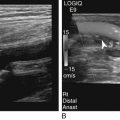
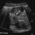
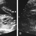
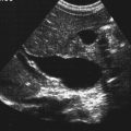
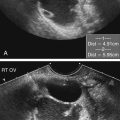
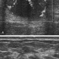
Charlotte Henningsen and Lennard D. Greenbaum • List the various causes of bleeding with pregnancy. • Differentiate the most common causes of bleeding in the first trimester of pregnancy from the most common causes of bleeding in the second and third trimesters. • Explain the β human chorionic gonadotropin levels that are used in diagnosis of ectopic pregnancy, gestational trophoblastic disease, and pregnancy failure. • List the causes of bleeding in the second and third trimesters of pregnancy, and differentiate the sonographic findings. • Identify Doppler findings seen with various causes of bleeding with pregnancy. • Describe the patient symptoms and sonographic findings associated with retained products of conception. Subchorionic hemorrhages are low-pressure hemorrhages that occur most commonly in the first trimester of pregnancy. They often result from implantation of the fertilized ovum into the uterus. These areas of hemorrhage are seen between the uterine wall and the membranes and are not associated with the placenta, which distinguishes subchorionic hemorrhage from abruptio placentae. Patients may have spotting or bleeding with or without uterine contractions. Subchorionic hemorrhage may spontaneously regress or may lead to spontaneous abortion (SAB); the prognosis is favorable when the fetal heartbeat is identified in the presence of a small hemorrhage. In addition, hemorrhage in the lower uterine segment has a better prognosis than hemorrhage located at the uterine fundus.1 Patients who present with bleeding in whom a subchorionic hemorrhage is found have a higher incidence of pregnancy loss than patients with a subchorionic hemorrhage without bleeding.2 Sonographic examination of a subchorionic hemorrhage reveals blood, which may initially be echogenic and progressively become anechoic (Fig. 21-2), located adjacent to the gestational sac and at the margin of the placenta. The lack of vascularity identified with color Doppler can help differentiate hematoma from a neoplasm. When the hemorrhage is anechoic, it may resemble a second gestational sac. The incidence of SAB is considered high, occurring in 15% of women with a known pregnancy.3 Depending on the patient presentation, abortion may be classified as threatened, missed, or complete. Threatened abortion is characterized by bleeding without cervical dilation. Missed abortion is characterized as embryonic death without expulsion of the products of conception. SAB is classified as complete when there is expulsion of the products of conception. Pregnancy loss is often idiopathic but may occur in aneuploid fetuses or as the result of maternal endocrine or vascular disorders, anatomic factors, or immunologic disease. When the pregnancy reaches 12 weeks’ gestational age, the SAB rate decreases dramatically. Correlation between the serum β human chorionic gonadotropin (β-hCG) and the findings in the uterus can be used to confirm whether or not the sonographic milestones of a first-trimester pregnancy are met. The gestational sac may be identified at 4.5 weeks by last menstrual period and should grow about 1 mm per day; additionally, the embryo grows at a rate of 1 mm per day. The size of the gestational sac and the crown-rump length should be consistent with each other. With endovaginal technique, the yolk sac should be visualized by the time the mean sac diameter reaches 8 mm; anomalies of the size, shape, or echogenicity of the yolk sac are associated with a poor prognosis. An embryo should be identified when the gestational sac measures 25 mm, and cardiac motion in the embryo should be visualized when the embryo measures 5 mm.2 Anembryonic pregnancy should be considered when an empty gestational sac is seen at this point in the gestation (Fig. 21-3). Embryonic bradycardia, defined as less than 85 beats per minute in a gestation less than 7 weeks’ gestational age and less than 100 beats per minute from 7 weeks’ gestational age forward, is associated with a poor prognosis.2 Failure to meet any of the milestones in the first trimester of pregnancy can suggest a poor pregnancy outcome. The values assigned to these thresholds have changed over time, and the authors are aware that discussion is ongoing regarding increasing the current thresholds; the literature should be monitored for current standards. A primary role of sonography in the evaluation of pregnancy in the first trimester is confirmation of viability. If the crown-rump length measures 5 mm and cardiac activity is not seen, the diagnosis of embryonic death can be made with confidence.2 Failure to meet other first-trimester sonographic milestones should be noted. A subchorionic hemorrhage may also lead to pregnancy loss. Ultrasound may be used to follow bleeding with pregnancy to monitor viability, to confirm fetal death, and to determine when dilatation and curettage (D&C) is necessary. Ectopic pregnancy is defined as a pregnancy located outside of the normal location (Fig. 21-4). It occurs in 2:100 pregnancies in the United States4 and is a leading cause of maternal death in the first trimester. A history of ectopic pregnancy also affects the future fertility of a patient and increases the risk of a repeat ectopic pregnancy. In addition to a history of ectopic pregnancy, risk factors include a history of pelvic inflammatory disease, tubal surgery, maternal congenital anomalies, late primiparity, defective zygote, fertility treatments, and intrauterine device (IUD) usage. The increased usage of assisted reproductive technology not only has increased the incidence rates of ectopic pregnancies and multiple gestations but also has increased the incidence rate of heterotopic pregnancies. The most common location of an ectopic pregnancy is in the fallopian tube, with a reported incidence 75% to 80% of ectopic pregnancies specifically in the ampullary portion.4 Ectopic pregnancies have also been identified in the cervix, ovary, uterine cornu, broad ligament, and abdomen. Interstitial or cornual pregnancy occurs in 2% to 5% of ectopic pregnancies.5 This type of ectopic pregnancy is located in the interstitial portion of the fallopian tube, which is located in the wall of the uterine cornu, and may result in massive hemorrhage, with rupture leading to serious complications, possible hysterectomy, and death. Ovarian pregnancies occur in 2% of ectopic pregnancies and may be indistinguishable from other ovarian pathologies.6 Cervical pregnancies are rare but can lead to massive hemorrhage that may necessitate hysterectomy to stop the bleeding. Cervical pregnancy may have an ultrasound appearance that is similar to a SAB because a gestational sac is identified within the cervix. Abdominal pregnancies also are rare and usually occur in the pelvis, although implantations have been reported in the upper abdomen, including in the liver. With the increasing frequency of cesarean section, ectopic pregnancies are also occurring in cesarean section scars. Heterotopic pregnancy (see Fig. 21-4, D-F) is defined as an intrauterine pregnancy (IUP) with an ectopic pregnancy. This type of ectopic pregnancy is rare, occurring in 1:10,000 to 1:50,000 spontaneous pregnancies but is relatively common in patients undergoing fertility treatment.6 The clinical symptoms of ectopic pregnancy include bleeding, pain, and a palpable adnexal mass. These symptoms are nonspecific and may be seen in patients who are not pregnant. In addition, women with an ectopic pregnancy may be asymptomatic or have symptoms more suggestive of a threatened abortion. The ultrasound findings paired with quantitative β-hCG levels assist clinicians in confirmation of an IUP or assessment of the risk of an ectopic pregnancy. The discriminatory zone defines the point at which a normal pregnancy should be identified sonographically, and the values may vary from institution to institution; additionally, these values vary depending on imaging technique. An intrauterine gestational sac should be identified in the uterus when scanning endovaginally if the β-hCG level is 1500 IU/L or greater.6 The absence of an IUP in the presence of these β-hCG levels suggests an increased risk of ectopic pregnancy. With normal pregnancy, the β-hCG levels should double every 2 days until they plateau. However, in the presence of an ectopic pregnancy, the levels increase more slowly, although a nonviable pregnancy could also be seen with this finding. The most specific sonographic finding diagnostic of ectopic pregnancy is an extrauterine gestational sac that contains a living embryo (see Fig. 21-4, C). Likewise, ectopic pregnancy is generally excluded in the presence of a living IUP, although heterotopic pregnancy should be considered in patients undergoing assisted reproductive technology. In the absence of an extrauterine embryo, other positive sonographic findings in combination or isolation may suggest ectopic pregnancy. The uterus may be consistent with a normal nongravid uterus or may have a decidual reaction or pseudogestational sac (see Fig. 21-4, A). A pseudogestational sac does not show the low-resistance, peritrophoblastic flow with Doppler that accompanies a normal IUP. A pseudogestational sac also lacks the “double sac” sign that represents the chorionic villi and endometrial cavity seen with early IUPs. The adnexa also should be carefully evaluated for signs of ectopic pregnancy. A live extrauterine pregnancy is infrequently identified, but endovaginal sonographic evaluation is able to identify findings suggestive of ectopic pregnancy with confidence. Studies have shown that the correct detection of tubal ectopias specifically has an overall sensitivity of 87% to 99%.7,8 An echogenic adnexal ring (see Fig. 21-4, F) may be seen with or without the presence of a yolk sac. A peritrophoblastic flow pattern may aid in distinguishing the adnexal ring from an ovarian cyst. However, a corpus luteum may also show a similar flow pattern. A hematosalpinx may be identified if the fallopian tube is distended with blood. An ectopic pregnancy may also be seen as a complex mass (see Fig. 21-18, B). A thorough investigation may reveal an empty uterus and normal-appearing adnexa. Identification of cul-de-sac fluid (see Fig. 21-4, G) is suggestive of ectopic pregnancy, whether or not other positive sonographic findings are present. Hemoperitoneum may appear anechoic, complex, or echogenic. When ultrasound does not show an IUP or a finding suggestive of ectopic pregnancy, clinical correlation determines when surgical, medical, or expectant management is appropriate. Because of the availability of sonography, most molar pregnancies are now identified in the first trimester before clinical signs are evident. In these incidences, sonography may suggest a missed abortion, and the diagnosis of molar pregnancy is confirmed by pathologic examination.9 When symptoms are present, they most commonly include vaginal bleeding; this may prompt evaluation of β-hCG, which is markedly elevated (>100,000 IU/mL9) in the presence of molar pregnancy. Maternal serum alpha fetoprotein levels are markedly low in pregnancies complicated by a complete hydatidiform mole. In addition to abnormal laboratory findings, the patient may have symptoms of hyperemesis, preeclampsia, thyrotoxicosis, or respiratory distress. Clinical examination may reveal a uterus that is greater in size than the expected gestational age and bilateral ovarian enlargement owing to theca-lutein cysts. A complete hydatidiform mole appears as an echogenic mass within the uterus that contains multiple cystic areas (representing hydropic chorionic villi) and that has been described as having a “snowstorm” appearance (Fig. 21-5, A). In addition, no fetus or fetal parts are identified. Early first-trimester findings may be nonspecific, simulating a missed abortion or anembryonic pregnancy, or may appear as an echogenic mass within the uterus. A complete hydatidiform mole with a coexistent fetus (see Fig. 21-5, C) is seen as a normal fetus and placenta with a concurrent molar pregnancy. This entity can be missed in the first trimester because of the presence of a live fetus, and the abnormal echogenicity of the placenta can be mistaken for hemorrhage.10 Differentiation from a molar pregnancy is important because of the varying malignant potential between these two entities. Although a complete mole with coexistent fetus has the same malignant potential as a solitary hydatidiform mole, a partial mole has a significantly lower malignant potential. In addition to the findings within the uterus, hyperstimulation of the ovaries may occur as a result of the greatly increased β-hCG levels. Bilateral theca-lutein cysts (see Fig. 21-5, B) are commonly seen in molar pregnancy; however, they are not likely to be seen in the first trimester.11 These enlarged ovaries may rupture or torque, causing severe pain. The sonographic description of theca-lutein cysts includes enlarged ovaries that contain multiple large cysts, giving them a “soap-bubble” appearance.
Bleeding with Pregnancy
First-Trimester Bleeding
Subchorionic Hemorrhage
Sonographic Findings
Abortion
Sonographic Findings
Ectopic Pregnancy
Sonographic Findings
Gestational Trophoblastic Disease
Molar Pregnancy
Sonographic Findings
![]()
Stay updated, free articles. Join our Telegram channel

Full access? Get Clinical Tree


Bleeding with Pregnancy


