5
 THE RIGHT ATRIUM
The right atrium is located anteriorly and to the right of the left atrium. The posterior portion of the right atrium (sinus venarum) has a smooth wall and receives the superior and inferior venae cavae and the coronary sinus (Fig. 5-1). The anterior portion of the right atrium is lined with coarse muscle bundles, named pectinate muscle (Fig. 5-1). The smooth and coarse regions within the right atrium are separated by a ridge called the crista terminalis (Fig. 5-1), which runs inferiorly and parallel to the openings of the inferior and superior venae cavae. The inferior vena cava enters the right atrium at its lowest part, near the interatrial septum (Fig. 5-1). The Eustachian valve, which represents a flap of endocardium, is located at the opening of the inferior vena cava (Fig. 5-1). This valve serves a critical function in the fetus as it directs highly oxygenated blood originating from the ductus venosus within the inferior vena cava to the foramen ovale in a caudal-to-cranial flow pattern. On occasions, the Eustachian valve can be imaged on fetal echocardiography in a four-chamber-view plane that is posterior to the optimal plane, where it can be mistaken for the flap of the foramen ovale. The coronary sinus, which delivers venous return from the heart, runs across the atrioventricular groove posteriorly and enters the right atrium inferiorly in proximity to the atrial septum and the inferior vena cava opening (1) (Figs. 5-1 and 5-2). The opening of the coronary sinus is also guarded by a valve called the Thebesian valve (Fig. 5-1). The superior vena cava (SVC) enters the right atrium anteriorly and has no flap at its opening. The sinoatrial (SA) node is located on the epicardium of the right atrial wall superiorly, just below the SVC (Fig. 5-1). The atrioventricular (AV)
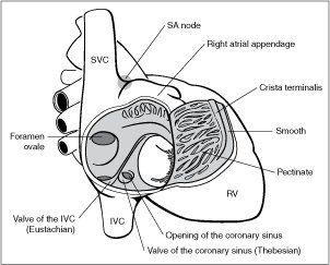
Figure 5-1. Right atrial internal anatomy. The posterior wall is smooth; the anterior wall is coarse. Note the openings of the inferior vena cava (IVC), superior vena cava (SVC), and coronary sinus. See text for details. RV, right ventricle; SA, sinoatrial.
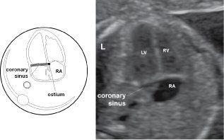
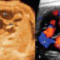
Figure 5-2. Transverse view of the fetal chest in a plane slightly caudal to the four-chamber-view plane showing the coronary sinus at the floor of the right atrium (RA). RV, right ventricle; LV, left ventricle; L, left.
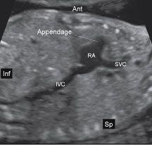
Figure 5-3. Parasagittal view of the right atrium (RA) with the superior vena cava (SVC) and inferior vena cava (IVC). Note the broad-shaped right atrial appendage, not seen at the four-chamber-view plane. Sp, spine; Inf, inferior; Ant, anterior.
node is located on the floor of the right atrium in proximity to the opening of the coronary sinus. The right atrial appendage is somewhat pyramidal in shape and has a broad base (Figs. 5-1 and 5-3). The Chiari network, another embryologic remnant, is composed of lacelike strands and is occasionally seen at the level of the tricuspid valve annulus within the right atrium. Table 5-1 lists the anatomic characteristics of the right atrium.
The two atria are separated by the atrial septum. The atrial septum is formed by the septum primum and secundum. The foramen ovale is formed as a perforation in the septum secundum (Fig. 5-4). The septum primum, embryologically the first atrial septum to develop, forms the leaflet of the foramen ovale. Atrial septal defects are categorized according to the
| TABLE 5-1 | Anatomic Characteristics of the Right Atrium |
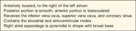
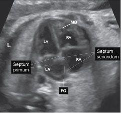
Figure 5-4. Apical four-chamber view of the fetal heart with the right atrium (RA), left atrium (LA), right ventricle (RV), and left ventricle (LV). At the apex of the heart in the right ventricle the typical thickened moderator band (MB) is recognized. Between both atria the interatrial septum is formed by both the septum primum and secundum. The flap of the foramen ovale is formed by the septum primum, while the foramen ovale (FO) is an opening in the midportion of the septum secundum. L, left.
anatomic location of the defect within the septum. Defects in the lower one third of the atrial septum are referred to as septum primum defects, defects in the middle one third are referred to as septum secundum defects, and defects in the posterior and superior portions of the septum are referred to as sinus venosus defects.
 THE TRICUSPID VALVE
The tricuspid valve prevents the blood from flowing back into the right atrium from the right ventricle during ventricular systole. This valve has three leaflets—anterior, septal, and posterior—named based on their orientation within the right ventricle. The valves are anchored by chordae tendineae, which prevents the leaflets from prolapsing into the right atrium during systole. Chordae tendineae insert into three papillary muscles. The anterior papillary muscle, the largest of the three and commonly seen on ultrasound, is located in the apex of the right ventricle and receives chordae tendineae insertions from the anterior and posterior leaflets (Fig. 5-5). The posterior papillary muscle is located in the posterior lateral wall and receives
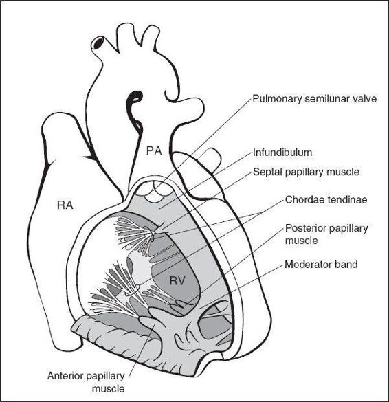
Figure 5-5. Internal anatomy of the right ventricle (RV), which is made up of an inlet, apical, and outlet portion. The tricuspid valve is made up of three leaflets and three papillary muscles. The moderator band occupies the apex of the RV. See text for details. RA, right atrium; PA, pulmonary artery.
Related posts:
Stay updated, free articles. Join our Telegram channel

Full access? Get Clinical Tree


