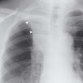
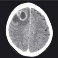
An ‘air bronchogram’ (arrow) can be seen clearly within the area of shadowing in the right middle zone. This shadowing has a distinct inferior margin (arrowheads) running along the line of the horizontal fissure which is the inferior boundary of the right upper lobe. This is therefore right upper lobe consolidation
11.2 Atelectasis

This patient was recovering from a widespread pneumonia when this CXR was taken. There are multiple bands of atelectasis (arrowheads) which are the result of the inflammatory process. There is a persistent area of consolidation in the region of the right hilum (*)
11.3 Bronchiectasis

Bronchiectasis is a difficult diagnosis to make with any great certainty on plain CXR imaging. The diagnosis is made accurately on high resolution CT (see Chapter 33). This CXR demonstrates ‘tram tracks’ (arrowheads) which represent thick-walled dilated bronchi
11.4 COPD

These lungs are hyperinflated as shown by the flattening of the hemidiaphragms (arrowheads). The anterior portion of more than seven ribs can be seen lying above the hemidiaphragms in the midclavicular line on both sides. The lungs are so hyperinflated that the inferior border of the heart is lifted away from the diaphragm
Consolidation
Consolidation is a pathological process caused by filling of the alveolar airspaces with fluid or debris such as:
- Inflammatory exudates, e.g. pneumonia (see Chapter 33).
- Haemorrhage, e.g. trauma, vasculitis, or pulmonary embolism.
- Transudates from various causes, e.g. cardiogenic, nephrogenic, neurogenic, ARDS and drug reactions.
- Secretions, e.g. alveolar proteinosis and mucus.
- Malignancy, e.g. bronchioalveolar cell carcinoma and lymphoma.
Consolidation appears as a ‘fluffy’ light grey density that is not well demarcated unless bound by a pleural margin, e.g. a fissure or lung edge. The classic signs are air bronchograms
Stay updated, free articles. Join our Telegram channel

Full access? Get Clinical Tree


