Michael M. Maher, Owen J. O’Connor
Image-Guided Drainage Techniques
Image-guided drainage is an established technique with a multitude of applications. The indications, techniques and management of image-guided catheter drainage, however, continue to evolve. This chapter provides an overview of the principles of image-guided drainage. We also discuss important technical aspects of specific drainage procedures, how to care for a drainage catheter and potential complications that can arise.
Indications and Contraindications
As a general rule, image-guided drainage is indicated for treatment of an accessible collection in a suitable patient that does not require immediate surgical intervention, to obtain a fluid sample for diagnostic purposes, to relieve symptoms or to inject a sclerosant.1 Image-guided drainage alone is sometimes sufficient for treatment of a collection, but it can also act as an adjunct or temporising measure before definitive surgical treatment.2 Drainage of a symptomatic collection such as an abscess is performed in order to drain pus from the cavity, working in conjunction with antibiotics. Infected collections accumulate antibiotics to a limited extent, which generally precludes effective treatment with antibiotics alone unless the collection is very small (1–3 cm).3 Antibiotic coverage is necessary for many drainage procedures, even if the collection is not infected, to reduce the chance of secondarily infecting the collection. Examples of non-infected symptomatic collections that often require drainage include hydronephrosis caused by ureteric obstruction, bowel obstruction or postoperative seroma, urinoma and haematoma. Diagnostic fluid samples help determine whether a collection is infected and may also help identify the source of a collection. Fluid should be analysed by Gram stain; culture and sensitivity analysis should also be performed. Analysis of the cell count is also useful for quantifying the number of white cells in a sample. Amylase, bilirubin, lymphocytes and creatinine content can be used to identify collections of pancreatic, biliary, lymphatic and urinary origins, respectively.4
There are few absolute contraindications to image-guided drainage. Profound uncorrected coagulopathy, clinical instability and lack of safe access to a collection are among the most common contraindications to image-guided drainage. In practice, many coagulopathies can be corrected to allow drainage. The authors stratify procedure-related bleeding risk into three categories: low, medium and high. Paracentesis, catheter exchange and aspiration are considered low risk. Abdominal and chest procedures are considered medium risk, whereas primary biliary or renal drainages are stratified as high risk. The authors consider correction of abnormal indices for a low-risk procedure if the platelet count is less than 30,000/µL and the international normalised ratio (INR) exceeds 2.5, or for a medium-risk procedure if the platelet count is less than 30,000/µL and the INR exceeds 1.7 or the partial thromboplastin time is 1.5 times normal. Correction is performed prior to a high-risk procedure if the platelet count is less than 50,000/µL and the INR exceeds 1.7 or the partial thromboplastin time is 1.5 times normal. These guidelines need to be tailored to the collection and the patient, taking into account the fact that an untreated abscess has a very high mortality rate, and successful drainage reduces morbidity.5,6 Encasement by bowel loops and large blood vessels preclude drainage catheter placement. A 19–22G needle can be used to traverse the small bowel for diagnostic sampling but risks infecting a fluid collection with enteric organisms. General anaesthetic or monitored anaesthesiology care is necessary for clinically unstable patients requiring image-guided drainage catheter placement. Lack of maturation of an abscess is a potential reason for close-interval observation before drainage. Peritonitis and a large volume of intraperitoneal air in the setting of a collection are indications for surgical rather than image-guided drainage. Air localised and contained within the vicinity of a collection, generally, does not preclude image-guided drainage, and is usually caused by local gastrointestinal perforation such as from appendicitis or diverticulitis. In cases where there is uncertainty, good communication between the interventional radiologist, referring physician and patient or family is indicated in order to reach consensus.
Imaging Guidance
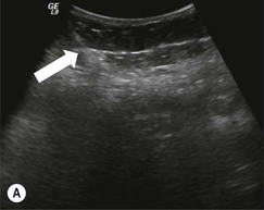
Ultrasound and CT are primarily used for image-guided drainage. Modern ultrasound provides excellent real-time visualisation of superficial structures, and good visualisation of viscera or collections. This facilitates careful monitoring of a catheter or needle as it is guided into a collection, irrespective of the plane of angulation. This is preferred in paediatric patients for whom radiation exposure should be minimised and can also be performed at the bedside in an intensive care unit (Fig. 88-1). It is important to maintain the needle or catheter in the plane of imaging when using ultrasound. If possible, a needle-probe angle of 55°–60° should be maintained to optimise reflection of the ultrasound beam and needle or catheter visualisation.7 The optimal grey-scale image map for ultrasound-guided procedures is different to that of diagnostic ultrasound, and should be sought. Additionally, frequency compound imaging is an ultrasonic imaging technology which can help with needle visualisation during drainage. Compound imaging emits ultrasound at multiple incident beam angles, which increases needle conspicuity because of increased artefact. Compound imaging reduces the pulse repetition frequency, however, which can cause image discontinuity. Ultrasound is often suboptimal for catheter guidance if a collection contains air or if there are bowel loops adjacent to a collection which prevent adequate imaging and increase the risk of bowel injury. Ultrasound guidance can be used in a hybrid manner for drainage purposes (Fig. 88-2). Combined with fluoroscopy, ultrasound can guide access and catheter placement into a large or partially visualised collection before optimal catheter manipulation and positioning by fluoroscopic guidance. Fluoroscopic guidance is also beneficial for guidance of catheters placed using the Seldinger technique and reduces the risk of losing access and kinking the guidewire (Fig. 88-2).
CT-guided fluoroscopy has many proponents since it offers potential real-time guidance and excellent spatial resolution, which can reduce procedural time. One study has shown a 37% reduction in needle placement time but no significant reduction in room time using CT fluoroscopy for interventional radiology procedures.8 CT fluoroscopy can be performed real time or by using a quick-check method. Real-time guidance involves holding the needle or catheter with a clamp and imaging as it is advanced. Quick-check CT guidance is used to image the needle tip after manipulation. Quick-check guidance considerably reduces fluoroscopic time and radiation dose compared with real-time guidance. Conventional CT guidance is favoured over ultrasound for drainage of deep collections with a difficult percutaneous access window.9 Superior spatial resolution of CT over ultrasound often allows better localisation of the margins of a collection, the thickness of the wall, the adjacent organs and the access route. Tilting the angle of the CT gantry in a cranial or caudal direction is a useful adjunct, which can help image a safe direct route of access into a collection that is not available in the axial plane. This can create an additional level of difficulty for the interventional radiologist; the gantry laser guide is very useful for catheter direction in this circumstance. Room time, available resources, user preference and experience have a determining impact on the choice of image guidance.
Patient Preparation and Care
The authors recommend broad-spectrum antibiotics at least 1 h prior to abscess drainage. This does not preclude culture of material from an abscess since the rind of tissue that surrounds an abscess excludes most abscesses from the normal circulation.
Written informed consent is an important aspect of image-guided drainage. Optimal informed consent entails description of the indications for image-guided drainage, the alternatives, the procedure itself, potential complications and also the expected treatment plan after drainage. Since drainage catheters often remain in place for weeks, it is important that the patient be made aware of this so as to avoid unrealistic expectations. Effective teamwork and open communication between all those involved in the patient’s care helps to reduce the risk of error and improves patient safety.10,11 Normally, nursing sedation and continuous monitoring of vital signs is required for image-guided drainage. Patients should fast for 8 h prior to conscious sedation. The authors normally use fentanyl citrate (Elkins-Sinn, Cherry Hill, NJ, USA) and midazolam (Versed; Hoffmann-La Roche, Nutley, NJ), supplemented by antiemetics where appropriate. General anaesthesia is required for paediatric patients and severely ill or uncooperative patients. The authors advocate 4 h close observation after drainage to assess for complications.
Catheter Insertion
The following paragraphs provide an overview of techniques used for generic image-guided drainage. Drainage procedures which require special techniques or consideration will be discussed later in the chapter. Percutaneous aspiration is less likely to treat an abscess adequately compared with drain insertion. A collection which communicates with the bowel, biliary or urinary tracts should not be treated by aspiration. Occasionally, a small collection inaccessible to drain insertion, such as an interloop abscess in an immunosuppressed patient with Crohn’s disease, may be treated by aspiration; otherwise, drain insertion is favoured.
CT-guided catheter placement is generally performed by one of two methods: tandem-trochar or Seldinger. Tandem-trochar technique relies on the placement of a catheter containing a hollow stiffener and a diamond-pointed stylet, parallel to a guide needle into a collection (Fig. 88-3). Trochar catheters are available from 8 to 16Fr in size. The authors normally use a hydrophilic-coated Ultrathane catheter with a locking loop (Cook, Bloomington, IN, USA) for image-guided drainage. A CT examination, with a radio-opaque grid on the patient’s skin enables a safe route for needle and catheter placement to be chosen. The distance from the skin surface to the abscess is measured and a 20G guide needle of appropriate length chosen. The portion of the needle outside the skin needs to be long enough to guide the trajectory of the catheter. Following cleansing and local anaesthetic administration, the needle is placed through the skin into the collection and a sample obtained for culture. This sample also allows assessment of the viscosity of the collection but the collection should not be aspirated further until the catheter is placed. A 10–12Fr catheter is usually necessary if the contents are frank pus; an 8–10Fr catheter may be adequate for less viscous fluid. The distance from the skin to the contents of the collection should be marked on the catheter. Following a skin incision and tissue separation adjacent to the guide needle, the catheter is introduced parallel to the guide needle, to the level of the mark on the catheter. The catheter may then be advanced over the stiffener or the stiffener withdrawn and the retention pigtail formed.
Once adequate catheter position is confirmed, the contents of the collection are evacuated, the catheter is secured to the skin with an adhesive device and a drainage bag is attached. Catheter irrigation at the time of abscess drainage using normal saline can increase drainage yield, disrupt adhesions and improve healing time. However, the volume of normal saline injected must not exceed that of the fluid drained from the collection to prevent cavity distension and reduce the risk of bacteraemia. The tandem-trochar technique is fast and does not require serial dilatation, the metal stiffener affords good catheter directionality. It should be noted that some collections have tough fibrous walls which can deflect the catheter and a malpositioned catheter will generally need to be withdrawn and replaced.
The Seldinger technique allows more controlled catheter placement, especially if there is high risk of catheter transgression of the posterior wall of a collection (Fig. 88-4) and can facilitate better drainage of large multiloculated collections by placement of a multi-sidehole catheter. A 19G ultrathin needle containing a stylet or a sheathed needle is initially placed into the collection. An 0.035-inch guidewire is advanced through the needle or sheath and, once adequate positioning is confirmed, the tract is serially dilated. The Seldinger technique is time consuming and tract dilatation can be painful, especially when traversing muscles. Dilatation also carries increased risk of content spillage during dilator exchange and before catheter placement. Manipulation of a dilator over the wire in a confined space also carries a risk of buckling the guidewire, which can hinder catheter placement.
Ultrasound-guided catheter insertion is performed under direct guidance following skin preparation (Fig. 88-1). A guide needle is not necessary unless one wishes to sample contents of the collection to assess consistency before choosing the catheter size.
Catheter Management
Drainage catheters should be flushed with normal saline every 8–12 h to maintain catheter patency and optimise drainage. A flush volume of 5 cc towards the patient and 5 cc towards the drainage bag via a three-way stopcock is normally sufficient unless a collection is very small or very large. Daily catheter outputs should be monitored and the contents of the drainage bag noted. This may alert one to evolving issues such as fistulisation or bleeding. In addition, difficulty flushing the catheter, pain on flushing and catheter withdrawal can signal blockage or displacement. Contrast injection into the catheter under fluoroscopic guidance is generally indicated in these circumstances. Catheter removal is considered in a well patient when daily outputs are low, normally on the order of 10 cc or less per day. Before catheter removal it is normally necessary to confirm complete drainage of the collection by CT or ultrasound imaging and confirm that the cavity has collapsed around the catheter and that there is no fistula present by fluoroscopic-guided injection. Catheter removal at this stage should be followed by complete collapse of the cavity. Optimal drainage and complication avoidance are best achieved by active participation of the interventional radiologist in patient rounds.12
Specific Drainage Techniques
Many image-guided drainages require technical modifications and special consideration. In this section we will discuss pertinent aspects of image-guided drainage procedures of the chest, liver, biliary system, pancreas, gallbladder, urinary tract, gastrointestinal tract, spleen, subphrenic region, peritoneum and deep pelvic territory, and organ traversal for drainage.
Chest
There are many indications for image-guided drainage in the chest, including pleural disease, lung parenchymal, pericardial and mediastinal collections. Pleural collections represent a common clinical problem for which image-guided drainage is recommended to reduce complications encountered as a result of blind drainage.13 Many types of pleural collections exist, and although diagnostic imaging is helpful, aspiration and drainage are often required for treatment and diagnosis. Pleural collections include effusions, haemothorax, empyema and pneumothorax. The success of image-guided drainage depends to a large extent on the contents of a pleural collection. It is, therefore, important for treatment decisions, to characterise the collection at the time of drainage, using biochemical, cytological and microbiological means. Effusions may be transudative or exudative. Light’s criteria are 98% sensitive and 80% specific for an exudative effusion if the ratio of pleural fluid protein to serum protein is greater than 0.5, if the ratio of pleural fluid lactate dehydrogenase (LDH) to serum LDH is greater than 0.6 or if the pleural fluid LDH is greater than  that of the normal upper limit for serum LDH.14
The natural history of an exudative pleural effusion is to evolve from free-flowing fluid to fibrinopurulent material, and later develop an organised fibrous pleural peel. Drainage of an exudative pleural collection should be performed early to prevent progression. Similarly, an infected pleural fluid collection should be drained as early as feasible in order to remove infection, sterilise the cavity, obliterate the pleural space and promote lung re-expansion, which improves drainage of secretions and return of pleural elasticity. Approximately 20% of patients with pneumonia develop an effusion. Since Light’s criteria are only reasonably specific for an exudative effusion, confirmation that the effusion is not a transudate is required in cases where there is discrepancy between the clinical and biochemical data. Fluid pH can be useful in these circumstances. The pH of an effusion is a strong predictor of the need for chest tube placement. Based on data from a meta-analysis, a pH less than 7.2 is an indication for chest tube placement.15
Image-guided pleural drainage catheters are smaller than surgical drains, measuring up to 16Fr in size, which means they are often better tolerated by patients. A smaller catheter is acceptable for treatment of free-flowing fluid but a large catheter is necessary for a complicated collection. Haemothorax caused by trauma is better treated using surgical drains (36–38Fr), although drainage of small blood-containing postoperative pleural collections may be attempted under imaging guidance. Ultrasound guidance is adequate for uncomplicated collections, but CT is usually needed for drainage of multiloculated pleural collections (Fig. 88-5). It is recommended that, where possible, the dependent portion of the collection be accessed, just above the adjacent rib, away from the paraspinal region where the neurovascular bundle lies lower in the intercostal space, and care should be taken to avoid insertion close to the scapula.16 The authors favour suturing chest tubes to the skin in order to avoid displacement. Coughing is common as the lung re-expands. For transudative effusions thoracentesis is often preferred over chest tube placement. Aspiration is stopped and the catheter withdrawn when 1.5 L of fluid is aspirated or the patient cannot tolerate further drainage. Imaging is always performed after pleural drainage to assess response and to check for pulmonary oedema and presence of pneumothorax. Daily chest radiographs are required and interval CT is indicated for assessment of pleural collections treated with a chest tube. It is also important to be aware of a vacuthorax phenomenon after pleural drainage so as to avoid unnecessary patient anxiety and harm.17 This manifests as a pneumothorax with or without fluid on imaging following pleural drainage. Patient’s generally have no symptoms from the pneumothorax and usually have an underlying diagnosis of chest malignancy. It is thought that inadequate surfactant causes non-compliance of the lung or the presence of restrictive pleural disease precipitates an asymptomatic hydropneumothorax in this setting, for which chest tube insertion is seldom required. Chest tubes inserted for parapneumonic and complicated effusions often do not drain large quantities of fluid immediately: 20 cm H2O suction is normally sufficient and a closed underwater seal system is required.
Chest tube removal is considered when there is clinical improvement, improved imaging appearances and absence of bubbling in the one-way valve system (suggests bronchopleural fistula), and outputs have dropped below 1–1.5 cc/kg body weight per day. A chest tube placed for pneumothorax can be removed 24 h after the pneumothorax has resolved. Modified drain management is often necessary as part of pleural collection treatment. For example, air leakage from a drain should prompt search for faulty connections or exposed catheter sideholes; otherwise, a bronchopleural fistula should be suspected. The degree of suction may be carefully reduced to deter further leakage.
Stay updated, free articles. Join our Telegram channel

Full access? Get Clinical Tree


