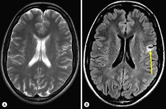
Alex Rovira, Pia C. Sundgren, Massimo Gallucci Idiopathic inflammatory-demyelinating diseases (IIDDs) represent a broad spectrum of central nervous system disorders that can be differentiated on the basis of severity, clinical course and lesion distribution, as well as imaging, laboratory and pathological findings. The spectrum includes monophasic, multiphasic and progressive disorders, ranging from highly localised forms to multifocal or diffuse variants. Relapsing-remitting and secondary progressive multiple sclerosis (MS) are the two most common forms of IIDDs.1 MS can also have a progressive course from onset (primary progressive and progressive relapsing MS). Fulminant forms of IIDDs include a variety of disorders that have in common the severity of the clinical symptoms, an acute clinical course and atypical findings on MR imaging. The classic fulminant IIDD is Marburg’s disease. Baló’s concentric sclerosis, Schilder’s disease and acute disseminated encephalomyelitis can also present with acute and severe attacks. Some IIDDs have a restricted topographical distribution, such as Devic’s neuromyelitis optica, which can have a monophasic or, more frequently, a relapsing course. Other types of IIDDs occasionally present as a focal lesion that may be clinically and radiographically indistinguishable from a brain tumour. It is difficult to classify these tumefactive or pseudotumoural lesions within the spectrum of IIDDs. Some cases have a monophasic, self-limited course, while in others the tumefactive plaque is the first manifestation or appears during a typical relapsing form of MS. MR imaging of the brain and spine is the imaging technique of choice for diagnosing these disorders, and, together with the clinical and laboratory findings, can accurately classify them.2 MS is a chronic, persistent inflammatory-demyelinating disease of the central nervous system (CNS), characterised pathologically by areas of inflammation, demyelination, axonal loss and gliosis scattered throughout the CNS. MS has a predilection for the optic nerves, brainstem, spinal cord and cerebellar and periventricular white matter. MS is one of the most common neurological disorders and the second cause of disability in Western countries in young adults of Caucasian origin. It is relatively common in Europe, the United States, Canada, New Zealand and parts of Australia, but rare in Asia, and in the tropics and subtropics of all continents. Multiple sclerosis is twice as common in women as in men; men have a tendency for later disease onset, with a poorer prognosis. The incidence of MS is low in childhood, increases rapidly after the age of 18, reaches a peak between 25 and 35, and then slowly declines, becoming rare at 50 and older.3 The aetiology of MS is still unknown, but it most likely results from an interplay between as-yet unidentified environmental factors and susceptibility genes. The clinical course of MS can follow different patterns over time, but is usually characterised by acute episodes of worsening (relapses, bouts), gradual progressive deterioration of neurological function, or a combination of both these features (relapsing MS). In a relatively small percentage of patients, the disease has a progressive course from onset, without acute relapses (primary progressive MS). Relapsing MS accounts for 85% of all MS. This clinical form typically presents as an acute clinically isolated syndrome attributable to a monofocal or multifocal CNS demyelinating lesion. The presenting lesion usually affects the optic nerve (optic neuritis), spinal cord (acute transverse myelitis), brainstem (typically an internuclear ophthalmoparesis) and cerebellum (clumsiness and gait ataxia). Over the following years, patients usually experience episodes of acute worsening of neurological function, followed by variably complete recovery (relapsing-remitting (RR) course). Clinical and subclinical activity is frequent in this form. After several years of the RR course, more than 50% of untreated patients will develop progressive disability with or without occasional relapses, minor remissions and plateaus (secondary progressive (SP) course).1 As long as the aetiology of MS remains unknown, causal therapy and effective prevention are not possible. Immunomodulatory drugs such as beta-interferon, glatiramer acetate, natalizumab and fingolimod can alter the course of the disease, particularly in the RR form, by reducing the number of relapses and the accumulation of lesions as seen on MR imaging, and by influencing the impact of the disease on disability. Patients with the SP form of MS, continuing relapses of activity and pronounced progression of disability may also benefit from immunomodulatory or immunosuppressive therapy. Primary progressive forms (PPMS) comprise approximately 10% of MS cases. This form of MS begins as a progressive disease with occasional plateaus and relapses, and temporary minor improvements. Progressive-relapsing MS follows a progressive course like PPMS, but shows clear acute relapses that may or may not be followed by full recovery.1 Compared to patients with the more frequent relapsing forms of MS, patients with PPMS have smaller T2 lesion loads, smaller T2 lesions, slower rates of new lesion formation and minimal gadolinium enhancement on brain MRI, despite their accumulating disability. The presence of extensive cortical damage, diffuse white matter tissue damage and prevalent involvement of the spinal cord may partially explain this discrepancy between the MR abnormalities and the severity of the clinical disease. Because patients with PPMS may have less inflammation than those with relapsing MS, they may be less likely to respond to immunomodulatory therapies. MR imaging is the most sensitive imaging technique for detecting MS plaques throughout the brain and spinal cord. Proton density (PD) or T2-weighted MR images show areas of high signal intensity in the periventricular white matter in 98% of MS patients. MS plaques are generally round to ovoid in shape and range from a few millimetres to more than 1 cm in diameter. They are typically discrete and focal at the early stages of the disease, but become confluent as the disease progresses, particularly in the posterior hemispheric periventricular white matter (Fig. 64-1). MS plaques tend to affect the deep white matter rather than the subcortical white matter, whereas small vessel ischaemic lesions tend to involve the subcortical white matter more than the periventricular white matter.4,5 The total T2 lesion volume of the brain increases by approximately 5 to 10% each year in the relapsing forms of MS.6 Both acute and chronic MS plaques appear bright on PD- and T2-weighted sequences, reflecting their increased tissue water content. The signal increase indicates oedema, inflammation, demyelination, reactive gliosis and/or axonal loss in proportions that differ from lesion to lesion. The vast majority of MS patients have at least one ovoid periventricular lesion, whose major axis is oriented perpendicular to the outer surface of the lateral ventricles (Fig. 64-2). The ovoid shape and perpendicular orientation derive from the perivenular location of the demyelinating plaques (Dawsons’ fingers). Multiple sclerosis lesions tend to affect specific regions of the brain, including the periventricular white matter situated superolateral to the lateral angles of the ventricles, the callososeptal interface along the inferior surface of the corpus callosum, the cortico-juxtacortical regions, and the infratentorial regions. Focal involvement of the periventricular white matter in the anterior temporal lobes is typical for MS and rarely seen in other white matter disorders (Fig. 64-3). The lesions commonly found at the callososeptal interface are best depicted by sagittal fast-FLAIR images; so this sequence is highly recommended for diagnostic MR imaging studies (Fig. 64-4). Histopathological studies have shown that a substantial portion of the total brain lesion load in MS is located within the cerebral cortex. Presently available MR imaging techniques are not optimal for detecting cortical lesions because of poor contrast resolution between normal-appearing grey matter (NAGM) and the plaques in question, and because of the partial volume effects of the subarachnoid spaces and CSF surrounding the cortex. Cortical lesions are better visualised by 2D or 3D fast-FLAIR sequences and newer MR techniques such as 3D double inversion recovery (DIR) MR sequences which selectively suppress the signal from white matter and cerebrospinal fluid (Fig. 64-5).7 Juxtacortical lesions that involve the ‘U’ fibres are seen in two-thirds of patients with MS. They are a rather characteristic finding in early stages of the disease, and are best detected by fast-FLAIR (Fig. 64-6) sequences. Multiple sclerosis frequently affects the brainstem and cerebellum, leading to acute clinical syndromes, such as trigeminal neuralgia, internuclear ophthalmoplegia, vertigo and ataxia. Later on, chronic damage to the posterior fossa causes chronic disabling symptoms such as ataxia and oculomotor disturbances. Acute symptomatic lesions appear as well-defined, hyperintense focal lesions that enhance with contrast administration on T1-weighted images (Fig. 64-7). Posterior fossa lesions preferentially involve the floor of the fourth ventricle, the middle cerebellar peduncles and the brainstem. Most brainstem lesions are contiguous with the cisternal or ventricular cerebrospinal fluid spaces, and range from large confluent patches to solitary, well-delineated paramedian lesions or discrete ‘linings’ of the cerebrospinal fluid border zones. Predilection for these areas is a key feature that helps to identify MS plaques and to differentiate them from focal areas of ischaemic demyelination and infarction that preferentially involve the central pontine white matter. Because of their short acquisition time and greater sensitivity, PD- and T2-weighted fast spin-echo sequences are preferred over conventional spin-echo or fast-FLAIR sequences for detecting posterior fossa lesions. Approximately 10–20% of T2 hyperintensities are also visible on T1-weighted images as areas of low signal intensity compared with normal-appearing white matter. These so-called ‘T1 black holes’ have a different pathological substrate that depends, in part, on the lesion age. The hypointensity is present in up to 80% of recently formed lesions and probably represents marked oedema, with or without myelin destruction or axonal loss. In most cases the acute (or wet) ‘black holes’ become isointense within a few months as inflammatory activity abates, oedema resolves and reparative mechanisms like remyelination become active. Less than 40% evolve into persisting or chronic black holes,8 which correlate pathologically with the most severe demyelination and axonal loss, indicating areas of irreversible tissue damage. Chronic black holes are more frequent in patients with progressive disease than in those with RR disease (Fig. 64-8), and are more frequent in the supratentorial white matter as compared with the infratentorial white matter. They are rarely found in the spinal cord and optic nerves. MS lesions of the spinal cord resemble those in the brain. The lesions can be focal (single or multiple) or diffuse, and mainly affect the cervical cord segment. On sagittal images, the lesions characteristically have a cigar shape and rarely exceed two vertebral segments in length. On cross-section they typically occupy the lateral and posterior white matter columns, extend to involve the central grey matter, and rarely occupy more than one-half the cross-sectional area of the cord9 (Fig. 64-9). Acute spinal cord lesions can produce a mild-to-moderate mass effect with cord swelling and may show contrast enhancement. Active lesions are rarer in the spinal cord than the brain, and are almost always associated with new clinical symptoms. The prevalence of cord abnormalities is as high as 74–92% in established MS, and depends on the clinical phenotype of MS. In clinically isolated syndromes, the prevalence of spinal cord lesions is lower, particularly if there are no spinal cord symptoms. Nevertheless, asymptomatic cord lesions are found in 30–40% of patients with a clinically isolated syndrome. In relapsing-remitting MS, the spinal cord lesions are typically multifocal. In secondary progressive MS, the abnormalities are more extensive and diffuse and are commonly associated with spinal cord atrophy. In primary progressive MS, cord abnormalities are quite extensive as compared with brain abnormalities. This discrepancy may help to diagnose primary progressive MS in patients with few or no brain abnormalities. Longitudinal and cross-sectional MR studies have shown that the formation of new MS plaques is often associated with contrast enhancement, mainly in the acute and relapsing stages of the disease10 (Fig. 64-10). The gadolinium enhancement varies in size and shape, but usually lasts from a few days to weeks, although steroid treatment shortens this period. Incomplete ring enhancement on T1-weighted gadolinium-enhanced images, with the open border facing the grey matter of the cortex or basal ganglia, is a common finding in active MS plaques and is a helpful feature for distinguishing between inflammatory-demyelinating lesions and other focal lesions such as tumours or abscesses11 (Fig. 64-11). Focal enhancement can be detected before abnormalities appear on unenhanced T2-weighted images, and can reappear in chronic lesions with or without a concomitant increase in size. Although enhancing lesions also occur in clinically stable MS patients, their number is much greater when there is concomitant clinical activity. Contrast enhancement is a relatively good predictor of further enhancement and of subsequent accumulation of T2 lesions, but shows no (or weak) correlation with progression of disability and development of brain atrophy. In relapsing-remitting and secondary progressive MS, enhancement is more frequent during relapses and correlates well with clinical activity. For patients with primary progressive MS, serial T2-weighted studies show few new lesions and little or no enhancement with conventional doses of gadolinium, despite steady clinical deterioration.12 Contrast-enhanced T1-weighted images are routinely used in the study of MS to provide a measure of inflammatory activity in vivo. The technique detects disease activity 5–10 times more frequently than clinical evaluation of relapses, suggesting that most of the enhancing lesions are clinically silent. Subclinical disease activity with contrast-enhancing lesions is four to ten times less frequent in the spinal cord than the brain, a fact that may be partially explained by the large volume of brain as compared with spinal cord. High doses of gadolinium and a long post-injection delay can increase the detection of active spinal cord lesions. Optic neuritis (ON) can usually be diagnosed clinically. MR imaging is not necessary to confirm the diagnosis, unless there are atypical clinical features (e.g. no response to steroids, long-standing symptoms). In these situations, brain and optic nerve MR imaging should be performed to rule out an alternative diagnosis, such as a compressive lesion.13 Coronal fat-saturated T2-weighted images are the most sensitive MR technique for depicting signal abnormalities. Focal thickening of the affected optic nerve reflects demyelination and inflammation (Fig. 64-12), which may persist for long periods despite improvements in vision and visual-evoked potential findings. Intense optic nerve enhancement seen on fat-suppressed contrast-enhanced T1-weighted images is a consistent feature of acute ON (Fig. 64-12). The length of the enhancing optic nerve segment on axial images correlates with the severity of visual impairment, but does not predict the degree of visual recovery. In MS, signal abnormalities may also be seen in the absence of acute attacks of ON. Atrophy of the brain and spinal cord is an important part of MS pathology, and a clinically relevant component of disease progression.14 Although this process is more severe in the progressive forms of the disease, it may also occur early in the disease process (Fig. 64-8). In fact, early atrophy seems to predict subsequent development of physical disability better than do measures of lesion load. The aetiology of CNS atrophy is multifactorial and likely reflects demyelination, Wallerian degeneration, axonal loss and glial contraction. CNS atrophy, which involves both grey and white matter, is a progressive phenomenon that worsens with increasing disease duration, and progresses at a rate of 0.6–1.2% of brain loss per year. Quantitative measures of whole-brain atrophy, acquired by automated or semi-automated methods, display this progressive loss of brain tissue bulk in vivo in a sensitive and reproducible manner. Subcortical brain atrophy is particularly well correlated to neuropsychological impairment, which can be explained by a disruption of frontal-subcortical circuits. Spinal cord atrophy is better correlated with motor disability. Marburg’s disease (MD) (also termed malignant MS) is a rare, acute MS variant that occurs predominantly in young adults. It is characterised by a confusional state, headache, vomiting, gait unsteadiness and hemiparesis. This entity has a rapidly progressive course with frequent, severe relapses leading to death or severe disability within weeks to months, mainly from brainstem involvement, or mass effect with herniation. Most of the patients who survive subsequently develop a relapsing form of MS. Because MD is often preceded by a febrile illness, this disease may also be considered a fulminant form of acute disseminated encephalomyelitis, if has a monophasic course. Pathologically, Marburg’s lesions are more destructive than those of typical MS or acute disseminated encephalomyelitis and are characterised by massive macrophage infiltration, acute axonal injury and tissue necrosis. Despite the destructive nature of these lesions, areas of remyelination are often observed. In MD, MRI typically shows multiple focal T2 lesions of varying size, which may coalesce to form large white matter plaques disseminated throughout the hemispheric white matter and brainstem (Fig. 64-13). Mild-to-moderate perilesional oedema is often present and the lesions may show peripheral enhancement.2 A similar imaging pattern is also seen in acute disseminated encephalomyelitis. Schilder’s disease (SD) is a rare acute or subacute disorder that can be defined as a specific clinical-radiological presentation of IIDD. It commonly affects children and young adults. The clinical spectrum of SD includes psychiatric predominance, acute intracranial hypertension, intermittent exacerbations and progressive deterioration. Imaging studies show large ring-enhancing lesions involving both hemispheres, sometimes symmetrically, and located preferentially in the parieto-occipital regions. These large, focal demyelinating lesions can resemble a brain tumour, an abscess or even adrenoleucodystrophy. MR features that suggest possible SD include large and relatively symmetrical involvement of brain hemispheres, incomplete ring enhancement, minimal mass effect, restricted diffusivity and sparing of the brainstem (Fig. 64-14).2 Histopathologically, SD consistently shows well-demarcated demyelination and reactive gliosis with relative sparing of the axons. Microcystic changes and even frank cavitation can occur. The clinical and imaging findings usually show a dramatic response to steroids. Baló’s concentric sclerosis (BCS) is thought to be a rare, aggressive variant of MS that can lead to death in weeks to months. The pathological hallmarks of the disease are large demyelinated lesions showing a peculiar pattern of alternating layers of preserved and destroyed myelin. One possible explanation for the concentric alternating bands in this variant of MS may be that sublethal tissue injury is induced at the edge of the expanding lesion, which would then stimulate the expression of neuroprotective proteins to protect the rim of periplaque tissue from damage, thereby resulting in alternative layers of preserved and non-preserved myelinated tissue.15 These alternating bands can be identified with T2 MR imaging, which typically shows concentric hyperintense bands corresponding to areas of demyelination and gliosis, alternating with isointense bands corresponding to normal myelinated white matter (Figs. 64-15 and 64-16). This pattern can appear as multiple concentric layers (onion skin lesion), as a mosaic, or as a ‘floral’ configuration. The centre of the lesion usually shows no layering because of massive demyelination. Contrast enhancement and decreased diffusivity are frequent in the outer rings (inflammatory edge) of the lesion (Fig. 64-16). On MR imaging, this Baló pattern can be isolated, multiple or mixed with typical MS-like lesions. Although Baló’s concentric sclerosis was initially described as an acute, monophasic and rapidly fatal disease that resembled Marburg’s disease, large Baló-like lesions are frequently identified on MR imaging in patients with a classical acute or chronic MS disease course, or in acute disseminated encephalomyelitis, with a non-fatal course. Infrequently, IIDDs present as single or multiple focal lesions that can be clinically and radiographically indistinguishable from a brain tumour. This situation represents a diagnostic challenge, and may require biopsy for definitive diagnosis, despite the clinical suspicion of demyelination. Given the hypercellular nature of these lesions, however, even the biopsy specimen may resemble a brain tumour. Large reactive astrocytes with fragmented chromatin (Creutzfeldt–Peters cells) are often present. In some cases, pseudotumoural IIDDs are the first clinical and radiological manifestation of MS. More commonly, tumefactive demyelinating plaques affect patients with a known diagnosis of MS (Fig. 64-17). In rare cases, pseudotumoural IIDDs have a relapsing course, with single or multiple pseudotumoural lesions appearing over time in different locations. On CT or MR imaging the pseudotumoural plaques usually present as large, single or multiple focal lesions within the cerebral hemispheres. Clues that can help to differentiate these lesions from a brain tumour are the relatively minor mass effect and the presence of incomplete ring enhancement on gadolinium-enhanced T1-weighted images, with the open border facing the grey matter of the cortex or basal ganglia (Fig. 64-18),16 sometimes associated with a rim of peripheral hypointensity on T2-weighted sequences. Devic’s neuromyelitis optica (NMO) is an uncommon and topographically restricted form of IIDD that is best considered to be a distinct disease rather than a variant of MS. NMO is characterised by severe unilateral or bilateral optic neuritis and complete transverse myelitis, which occur simultaneously or sequentially within a varying period of time (weeks or years), without clinical involvement of other CNS regions. The incidence and prevalence of NMO are unknown, but the condition likely accounts for less than 1% of IIDDs in Caucasians. NMO affects females almost exclusively. Approximately 85% of patients have a relapsing course with severe acute exacerbations and poor recovery, accumulating increasing neurological impairment and a high risk of respiratory failure and death due to cervical myelitis.17 Clinical features alone are insufficient to diagnose NMO; CSF analysis and MR imaging are usually required to confidently exclude other disorders. Cerebrospinal fluid pleocytosis (>50 leucocytes/mm3) is often present, while CSF oligoclonal bands are seen less frequently (20–40%) than in MS patients (80–90%). A serum autoantibody marker for NMO (NMO-IgG) has been recently developed. The target antigen of NMO-IgG is aquaporin-4, a water channel located on the foot process of the astrocyte. It is associated with tight endothelial junctions and cerebral microvessels and plays a critical role in maintaining fluid homeostasis in the CNS. This autoantibody is reported to have a sensitivity of 73% and a specificity of 91% for NMO. It may be helpful for distinguishing this form of IIDD from MS and may predict relapse and conversion to NMO in patients presenting with a single attack of longitudinally extensive myelitis. Wingerchuk et al. have proposed a revised set of criteria for diagnosing NMO (Table 64-1).18 These new criteria remove the absolute restriction on CNS involvement beyond the optic nerves and spinal cord, allow any interval between the first events of optic neuritis and myelitis, and emphasise the specificity of longitudinally extensive spinal cord lesions on MR imaging and NMO-IgG seropositive status. Devic’s neuromyelitis optica is a B-cell-mediated disorder that can coexist with diverse systemic autoimmune diseases, such as systemic lupus erythematosus, Sjögren’s syndrome and autoimmune thyroiditis. The presence of prodromal factors such as fever, infections and autoimmune abnormalities suggest that previous infectious-inflammatory events may be involved in the pathogenesis of the disease.19 MR imaging of the spinal cord shows extensive cervical or thoracic tumefactive myelitis, involving more than three vertebral segments on sagittal and much of the cross-section on axial T2-weighted images, which sometimes enhance with gadolinium for several months (Fig. 64-19). In some cases, the spinal cord lesions are small at the onset of symptoms, mimicking those in MS, and then progress in extent over time. These lesions are usually located centrally, can progress to atrophy and necrosis, and may lead to syrinx-like cavities on T1-weighted images (Fig. 64-20
Inflammatory and Metabolic Disease
Idiopathic Inflammatory-Demyelinating Disorders of the Central Nervous System
Multiple Sclerosis
MR Imaging
Brain.
Multiple Sclerosis Variants
Marburg’s Disease
Schilder’s Disease
Baló’s Concentric Sclerosis
Tumefactive or Pseudotumoural IIDDs
Devic’s Neuromyelitis Optica
![]()
Stay updated, free articles. Join our Telegram channel

Full access? Get Clinical Tree


Inflammatory and Metabolic Disease
Chapter 64


