Clinical condition
Incidence per year
References
Prevalence
References
Distal biceps tendon pathology
1.2 per 100,000
Safran and Graham (2002)
–
Triceps tendon ruptures
<1 %
Anzel et al. (1959)
>70 cases reported in the literature
Harris et al. (2004)
Elbow dislocation
6 per 100,000
Eygendaal (2009)
–
Medial epicondylitis
1 per 1000
Of all musculoskeletal disorders, 0.4 %
Lateral epicondylitis
4–7 per 1,000
1–3 % in the general population
Radial head fractures
–
5 % of all fractures
Mason (1954)
19.2 Functional Anatomy
The elbow plays an important role in the flexion-extension of the arm and supination-pronation of the forearm. It consists of three bones: the distal part of the humerus and the proximal parts of the ulna and the radius. These three bones articulate in the elbow in three separate joints: the radiohumeral joint, the ulnohumeral joint, and the proximal radioulnar joint.
The three elbow joints are surrounded by a joint capsule. It covers the tip of the olecranon, the coronoid process, and radial fossa but not the humeral epicondyles. The capsule is most lax at 80° of flexion and holds a capacity of 23 ml in this position (van Glabbeek and Clockaerts 2009). Patients with acute elbow injury and inflammation find this position therefore most comfortable.
Elbow stability results from the interplay of the articular surfaces, ligaments, and muscles. The radial head has an important role in maintaining elbow stability. The three primary static stabilizers of the elbow are the ulnohumeral articulation and the medial and lateral collateral ligaments. Secondary constrains include the radial head, capsule, and the common flexor and extensor origins. The muscles around the elbow, especially the anconeus, triceps, and biceps, function as dynamic stabilizers. If the coronoid process or medial collateral ligament is injured, the radial head becomes a critical stabilizer (O’Driscoll et al. 2001). The radiohumeral joint is the primary restraint to proximal migration of the radius. The interosseous membrane, a fibrous membrane between radius and ulna, and the triangular fibrocartilage complex (TFCC) at the distal radioulnar joint also contribute to longitudinal stability of the forearm (Hotchkiss et al. 1989).
Normal range of motion (ROM) is from full extension of 0–145° of flexion. Pronation and supination show large normal variations but usually are 80° of pronation and 85° of supination. Interindividual variation is wide (Soucie et al. 2011). Full ROM is not necessary for normal activities of daily living. Morrey et al. showed that for most activities in daily life, flexion-extension of 130 to −30° and a pro-supination arc of 100° would be sufficient (Morrey et al. 1981).
19.3 Etiology and Injury Mechanism
The elbow facilitates the highly skilled functions of the forearm, wrist, and hand that are needed for athletic endeavors. Athletic activities involving throwing, catching, hammering, pushing, pulling, and hitting with a racket, stick, or club produce significant stresses around the elbow joint that can result in acute or chronic injuries. Injuries to the elbow may involve the muscles, ligaments, tendons, capsule, bones, articular surfaces, and nerves or a combination of these structures and subsequently impair elbow function. Elbow injuries, particularly chronic overuse injuries, occur most commonly in athletes that participate in throwing or overhead activities and frequently are the result of valgus stress (Safran 2009).
Injuries to the elbow may be the result of acute trauma, such as a fall on an outstretched hand or a direct injury to the elbow itself. This may result in fractures with or without dislocation of the elbow joint. The majority of injuries to the elbow in the athlete are chronic overuse injuries. These injuries are the result of repetitive intrinsic or extrinsic overload, or both, resulting in micro-rupture of soft tissue such as ligament or tendon. Intrinsic overload is the force from muscular contraction which can lead to tendinitis or muscular injury. Extrinsic overload is a tensile overload caused by excessive joint torque forces stressing the soft tissue which results in stretching and eventual disruption. Extrinsic overload may also be due to compression of the soft tissues causing abrasion or impingement of the tissue. These micro-ruptures of the soft tissue may result in compromise of the soft tissue if followed by an imperfect healing process. In children, the growth plates are most susceptible to stress injuries. This is important, because a forceful acute injury in a patient with open growth plates as well as microtrauma may result in an apophyseal avulsion.
During overhead athletic activities, tremendous valgus forces are generated across the medial side of the elbow joint, resulting in tensile forces on the medial site, compression forces on the lateral site and shear forces in the posterior compartment. This combination of symptoms is often referred to as “valgus extension overload” (Ahmad and ElAttrache 2004).
19.4 Sports Injuries
19.4.1 Anterior Elbow Soft Tissue Injuries
19.4.1.1 Distal Biceps Tendon Pathology
At its origin, the biceps consists of two heads, a short head originating from the coracoid process of the scapula and a long head originating from the superior lip of the glenoid. The short head inserts distally to the radial tuberosity and is believed to be a more powerful flexor of the elbow, whereas the tendon of the long head inserting on the tuberosity further from the axis of rotation of the forearm is believed to be a stronger supinator. The bicipital aponeurosis consists of three layers and completely encircles the ulnar forearm flexor muscles. This aponeurosis may be important in stabilizing the tendons distally. A bursa is a potential cavity lined by a synovial membrane, which lies between two adjacent structures, aiming to provide friction-free motion. The bicipitoradial (or cubital) bursa either fully or partially envelops the distal biceps tendon. The bursa can exist as a distinct structure or can communicate with the elbow joint or other bursae around the elbow. The distal biceps tendon can be inflamed or (partially) ruptured.
Distal biceps tendon ruptures make up 3–12 % of all biceps injuries (D’Alessandro et al. 1993). The dominant arm is affected 86 % of the time (Safran and Graham 2002). The average age of patients is 50 years old (range 18–72) (Ramsey 1999). Risk factors include male gender, smoking, anabolic steroid use, and bodybuilding (Visuri and Lindholm 1994). The proposed etiology includes the presence of a bony prominence or irregularity of the radial tuberosity, radial bursitis, a watershed area of poor arterial supply, and mechanical impingement of the tendon (Hughes and Morrey 2000). The tendon will most commonly tear at its insertion. Tears within the length of the tendon and at the musculotendinous junction are rare (Schamblin and Safran 2007). The proximal portion of the tendon is more likely to retract into the brachium if the bicipital aponeurosis is also torn.
The patient usually recounts having sustained a sudden, unexpected, sharp extension load to an elbow flexed at 90°, followed by sudden antecubital pain. The patient may complain of weakness and aching in supination and flexion of the elbow with physically demanding activities (Ramsey 1999). Immediately after the injury, antecubital ecchymosis may be visible on examination; the patient may feel tenderness over the radial tuberosity or have a palpable gap in their tendon. There is usually weakness of supination. With partial ruptures, there may be tenderness over the radial tuberosity with no palpable gap in the tendon but crepitus or grinding on rotation of the forearm (Dellaero and Mallon 2006). Complete rupture is suggested by a positive hook test. The patient flexes the elbow to 90° and actively supinates the forearm. The examiner then hooks their finger under the biceps tendon from the lateral side. With a complete rupture, the examiner is unable to hook their finger under the cordlike biceps tendon (O’Driscoll et al. 2007).
19.4.2 Posterior Elbow Soft Tissue Injuries
19.4.2.1 Olecranon Bursitis
Olecranon bursitis is the most common form of superficial bursitis. The olecranon bursa lies superficial to bone and provides frictionless motion of the skin over the bony prominence of the olecranon. Given its superficial location on the extensor aspect of the elbow, it is vulnerable to repetitive trauma and penetrating injury.
Septic olecranon bursitis is caused by direct inoculation through the overlying skin or by seeding from surrounding tissues or a distant site (Ho et al. 1978). It can also be a presentation of septic arthritis of the elbow in cases where there is communication between the bursa and the elbow joint, e.g., rheumatoid arthritis. It has been reported to account for approximately 20 % to one third of cases of acute bursitis (Stell 1996). Immune compromise plays an important role in septic olecranon bursitis with approximately half of the patients having coexisting conditions such as alcohol abuse, steroid use, diabetes mellitus, renal impairment malignancy, or others which can impair the patient’s immune function (Behar and Chertow 1998). Septic bursitis can be difficult to differentiate clinically from a sterile inflamed bursa. The diagnosis is confirmed by aspiration and positive fluid culture. The most common organism is staphylococcus aureus (80–90 %) with beta hemolytic streptococcus being the next most common (Zimmermann et al. 1995).
More common is sterile inflammatory bursitis. Overuse or repetitive stress can result in recurrent inflammation of the olecranon bursae and has been named “student’s elbow” or “miner’s elbow.” Rheumatoid disease, gout, chondrocalcinosis, hydroxyapatite crystal deposition, and pigmented villonodular synovitis have all been associated with olecranon bursitis (Mathews et al. 1981). The condition has also been seen in uremic patients, especially those undergoing dialysis (Irby et al. 1975). It has been reported to occur in athletes due to repetitive trauma after falls on artificial turf.
19.4.2.2 Triceps Tendinopathy
Triceps tendon ruptures are among the rarest of tendon injuries with an incidence of <1 % (Anzel et al. 1959). The male to female ratio is 3:2, with a mean age of 33 years and range of 7 to over 80 years (Viegas 1990). The mechanism of injury is typically that of an extending elbow that is suddenly decelerated. The triceps muscle pulls the distal attachment off of the olecranon of the ulna. The tear is most commonly at the insertion of the tendon. Tears of the musculotendinous junction or the muscle belly are very rare. It is not uncommon for the patient to suffer associated injures such as a fracture of radial head or wrist (Levy et al. 1982).
The patient will usually recount a fall onto an outstretched hand, possibly with an extending elbow forced into flexion. Pain is typical over the olecranon at the site of the insertion of the triceps, and tenderness is usually elicited on palpation. There may be a palpable defect in the tendon. There is usually weakness of elbow extension against resistance. The anconeus muscle can sometimes overcome this weakness with the aid of gravity; therefore, extension should be assessed by asking the patient to extend the elbow against gravity with the affected arm held over the head (Bain et al. 2009).
Another triceps tendinopathy is the “snapping tendonitis.” A snapping elbow is a sensation that can be caused by a portion of the triceps mechanism. It may be confused with subluxation of the ulnar nerve over the medial epicondyle of the humerus on flexion and extension of the elbow (Spinner and Goldner 1998). Snapping tendonitis is caused either by abnormal medial triceps insertion, an aberrant triceps tendon, or cubitus varus. Both snapping tendonitis and subluxation of the ulnar nerve typically present with concurrent irritation of the ulnar nerve (Rolfsen 1970).
19.4.2.3 Posteromedial Impingement
Posteromedial impingement can be seen as an isolated condition or as part of the valgus extension overload. It is the result of repetitive microtrauma in the elbow (Kooima et al. 2004). Increased forces are transmitted to the posteromedial aspect of the olecranon and olecranon fossa articulation. This is often a result of attenuated medial soft tissues, namely, the anterior bundle of the medial collateral ligament (MCL) (Ahmad and ElAttrache 2004). In addition to pain, patients may report locking or limited elbow extension from an olecranon osteophyte.
The physical exam can be highly suggestive of valgus extension overload. Pain will be reproduced with the valgus extension overload test, where the slightly flexed elbow is forced into extension while applying a valgus load.
Given the role of MCL deficiency in the progression of valgus extension overload, the elbow must be assessed for valgus instability from 30° to 70° of flexion, as the MCL plays the most prominent role in stability in 70° of flexion (Eygendaal and Safran 2006). It is always critical to compare the stability to that of the uninjured limb, and it has been shown that neutral forearm rotation is the best position in which to uncover valgus laxity. A number of specialized tests have been described to test the integrity of the MCL. The moving valgus stress test is deemed positive when pain is reproduced in the 80–120° range. Another common test is the modified milking maneuver, which should be performed at 70° of elbow flexion (see Fig. 19.1) (Safran et al. 2005).
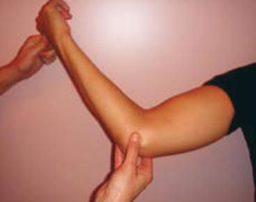
Fig. 19.1
Milking maneuver of the elbow
While the term posterior impingement has often been used to describe posteromedial impingement, true posterior impingement also exists as a discrete entity. It can stem from repetitive hyperextension microtrauma or emerge as the result of singular acute trauma. Due to its mechanism, it is seen most typically in football linemen, gymnasts, rodeo riders, weight lifters, fastpitch softball pitchers, and those competing in shot put (Moskal 2001). Patients typically complain of posterior pain, locking from loose bodies, or extension loss.
In true posterior impingement, instability should not be present. Instead, a block to terminal extension and pain with forced extension are often found. Imaging will often reveal direct posterior osteophytes. If posteromedial or posterolateral osteophytes are found instead, it should alert the physician to the possibility of coexisting valgus or varus laxity, respectively.
Posterolateral elbow impingement represents a heterogeneous group of bony and soft tissue impingement pathologies sharing the common absence of medial-sided instability.
19.4.3 Medial Elbow Soft Tissue Injuries
19.4.3.1 Medial Epicondylitis
Medial epicondylitis or golfer’s elbow is much rarer than its lateral counterpart. Of all epicondylitis diagnoses, only 10–20 % involves the medial side (Shiri et al. 2006). Medial epicondylitis occurs mostly in the fourth and fifth decades and affects men and women equally (O’Dwyer and Howie 1995). Medial epicondylitis is caused by forceful or repetitive activity (e.g., wrist flexion, forearm pronation) of the upper extremity (mostly occupational or sports related) and is also associated with smoking and obesity (Shiri et al. 2007). Commonly, the dominant arm is affected.
Repetitive overuse may lead to microtearing of the musculotendinous origin at the medial humeral epicondyle, mostly of the pronator teres, flexor carpi radialis, and palmaris longus muscles. Subsequent degeneration and failure of tendon healing (disruption of the normal collagen architecture with ingrowth of fibroblastic and granulation tissue) have been reported (Ollivierre et al. 1995). The pathology is degenerative rather than inflammatory; therefore, the term “tendinosis” is more appropriate (Nirschl and Ashman 2004).
A “burning” pain along the medial epicondyle, aggravated by repetitive wrist activity characterizes medial epicondylitis. The pain may radiate into the forearm. The onset of symptoms is usually insidious but may be acute – following an acute event. Range of motion is often unaltered but may be affected later on. Although articular signs are normal, grip strength may be decreased. In up to 10 % of all cases, involvement of the ulnar nerve is seen with numbness of the fourth and fifth fingers especially at night. Motor dysfunction of the ulnar nerve is rarely encountered. Just proximal to the medial epicondyle, the intermuscular septum inserts on the medial epicondyle; inflammation of this structure can mimic medial epicondylitis. In general, muscle function is less impaired in medial epicondylitis than in lateral epicondylitis (Pienimaki et al. 2002).
Medial epicondylitis may be diagnosed from the patient’s history and clinical examination. Palpation may reveal point tenderness directly on the medial epicondyle or slightly (up to 5 mm) distally and anterior to it. Resisted wrist flexion or pronation aggravates pain along the medial elbow. Grip strength is commonly diminished in the affected extremity.
However, the examiner should evaluate the cervical spine and shoulder as well in order to exclude radicular or referred arm pain.
19.4.3.2 Medial Collateral Ligament Injuries
In the normal elbow joint, stability is maintained by the combination of joint congruity, capsule-ligamentous integrity, and balanced intact musculature. The olecranon and olecranon fossa articulation provides primary stability at less than 20° of elbow flexion or flexion greater than 120°. In-between stability is provided by soft tissue constraints, mainly the MCL (Morrey et al. 1991).
The MCL consists of an anterior part or anterior medial collateral ligament (AMCL), a posterior part or posterior medial collateral ligament (PMCL), and a transverse band (see Fig. 19.2). This transverse band is also known as Coopers’ ligament and originates and inserts on the ulna and does not provide stability. Its function is unknown.

Fig. 19.2
Anatomy of the medial collateral ligament complex
The three most common causes of MCL injury are elbow dislocation, chronic attenuation in athletes, or acute valgus injury. The incidence of elbow dislocation in the general population is estimated to be 6/100,000. More than 95 % of all dislocations occur in a posterolateral direction. Posttraumatic chronic ligamentous instability used to be divided into medial or valgus instability and posterolateral rotatory instability (PLRI).
A new entity, the varus posteromedial injury (PMRI) mechanism, was more recently recognized and is increasingly understood. In PMRI, a fall onto the extended arm leads to a varus force with a posteromedial rotation. The varus force creates a lateral collateral ligament injury and a fracture of the anteromedial facet of the coronoid process, with resultant varus subluxation of the elbow that in some cases is apparent only on stress views of the elbow. If the injury force continues, the medial collateral ligament fails, and the elbow can dislocate completely. Complete dislocation and medial collateral ligament injury usually occur in association with a small anteromedial facet coronoid fracture, and larger fractures are associated with subluxation or disruption injuries rather than dislocation injuries. The radial head is not usually injured.
History taking is of utmost importance in the work-up of MCL insufficiency. At physical examination, comparison with the uninvolved elbow should always be performed to differentiate between physiologic laxity and pathologic instability. The degree of instability is often underestimated. Occasionally, the presenting symptoms of valgus extension overload (as posteromedial elbow pain, ulnar nerve symptoms, and chondromalacia at the lateral side) overshadow the symptoms of MCL insufficiency. Physical examination should also assess the degree of extension loss. The joint must be tested for valgus instability in 30° and 90° of flexion. Comparison with the uninvolved elbow should always be performed to differentiate between physiologic laxity and pathologic instability. The degree of instability is often underestimated.
Medial-sided pain of the elbow is not always a tendinosis of the flexor tendons or medial epicondylitis. Medial epicondylitis can be confused with and may coexist with MCL injury.
19.4.4 Lateral Elbow Soft Tissue Injuries
19.4.4.1 Lateral Epicondylitis
Lateral epicondylitis or tennis elbow is a common disorder in primary care. The name tennis elbow is derived from the description of “lawn tennis arm.” However, tennis contributes in only 5–10 % of all cases (Gruchow and Pelletier 1979). It is rather related to manually intensive work, requiring forceful and repetitive rotation of the forearm, wrist extension, or flexion (e.g., in mechanics, butchers, construction workers). Usually, the condition occurs with a peak between 35 and 54 years of age, and the dominant arm is involved (Hamilton 1986). Men and women are equally affected, according to most studies (Allander 1974).
Repetitive rotation causes degenerative inflammation of the wrist extensors that insert on the lateral epicondyle, most commonly the extensor carpi radialis brevis (ECRB). The pathology is degenerative rather than inflammatory; therefore, the term “tendinosis” is more appropriate (Nirschl and Ashman 2004).
The correct diagnosis of tennis elbow starts with a thorough history, revealing an activity or occupationally related “burning” pain located over the lateral epicondyle of the elbow or over the origin of the ECRB. The pain may radiate proximally or distally and is aggravated by lifting, gripping, or repetitive wrist activity. The onset of symptoms may be acute – secondary to an acute event –, or more gradual as a result of repetitive microtrauma. Although articular and neurological signs are normal, grip strength may be decreased. Eventually, even simple daily activities – such as shaking hands or turning a doorknob – may be painful. In severe cases, pain may also occur during the night or at rest.
Related posts:


Stay updated, free articles. Join our Telegram channel

Full access? Get Clinical Tree


