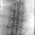
Fig. 19.1
Anterior view demonstrating the four parts of the parietal pleura
The line where the costal pleura becomes diaphragmatic pleura is called the costodiaphragmatic reflection. The line where costal pleura becomes mediastinal pleura is called the costomediastinal reflection. The right pleural reflection passes across the sternoclavicular joint and proceeds near the midline of the sternum, inferiorly from the level of the second rib to the 6th costal cartilage, swings laterally to cross the 8th rib in the midclavicular line, the 10th rib in the midaxillary line, and the 12th rib posteriorly, near the midline. The left pleural reflection is similar except at the 4th costal cartilage; the line swings laterally to the left border of the sternum. At the left and right costodiaphragmatic reflection, the pleura extends caudally without intervening lung tissue. This caudal extension forms a potential space (in the pleural cavity), the costodiaphragmatic recess. At the costomediastinal reflection, on the left side, lung tissue does not extend up to the costomediastinal reflection because of the cardiac notch in the left lung, and another potential space, the costomediastinal recess, is formed.
As these lines of reflection are fixed, intrapleural analgesia can technically be instituted anywhere with those boundaries.
Anteriorly, laterally, and posteriorly, the parietal pleura is in close approximation to the intercostal nerves. The parietal pleura has abundant sensory innervation from the phrenic and intercostal nerves (Fig. 19.2). Superiorly, the lower roots of the brachial plexus pass a short distance over the cupola before reaching the first rib. Medially, the sympathetic chain, splanchnic, phrenic, and vagus nerves are also adjacent. Because the epidural and subarachnoid spaces are further away, they are generally not thought to be involved during an intrapleural injection. However, since these spaces are only separated from the parietal pleura by fat and loose connective tissue of the epidural and paravertebral spaces, there may be tracking of anesthetic solution if there is a breach of the parietal pleura.

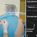
Fig. 19.2
Nerve supply of the thorax and intrapleural space
The visceral pleura is the innermost layer that is adherent to the substance of the lungs. It is continuous with the parietal pleura at the hilus of the lung. The intrapleural cavity is the potential space between the visceral and the parietal pleura. The space is 10–20 μm in width and has a static volume of 0.1–0.2 ml/kg. The micron-covered mesothelial surface of the parietal pleura facilitates the absorption of the local anesthetic and its diffusion into the intrapleural space. The pleura receives innervation from the phrenic and sympathetic nerves, and therefore, intrapleural administration of anesthetics may affect neural conduction on both types of nerves. Intrapleural analgesia can be accomplished by placing the anesthetic solution between the parietal and visceral pleurae, in this potential space (Fig. 19.3). Intrapleural analgesia can also be accomplished by placing a catheter deep to the internal intercostal muscle but superficial to the parietal pleura.

Fig. 19.3
Anatomy of the pleural cavity after (left) and before (right) injection of solution into the intrapleural space
The intercostal space of the posterior chest wall has three layers: the external intercostal muscle; the posterior intercostal membrane, which is the aponeurosis of the internal intercostal muscle; and the intercostalis intimus muscle, which is a continuation of the transversus abdominis. The intercostal nerves lie in between the posterior intercostal membrane and the intercostalis intimus. The intercostal intimus is incomplete and allows fluid to pass freely into the intrapleural space, whereas the posterior intercostal membrane forms a complete barrier beneath the external intercostal muscle.
Therefore, as previously stated, intrapleural analgesia can be accomplished in one of two ways. A catheter can be placed either deep to the internal intercostal muscle but superficial to the parietal and the visceral layers of the pleura or the catheter can be placed between the parietal and the visceral layers of the pleura. The local anesthetic can thus spread to adjacent intercostal nerves and paravertebral nerves, but spread to the epidural or subarachnoid space does not usually occur. Local anesthetic that is placed in the intrapleural space will diffuse out to anesthetize the thoracic somatic and lower cervical and thoracic sympathetic nerves that lie close to the parietal pleura.
Indications
Intrapleural analgesia can be used in the management of pain for the upper extremity, chest wall, thoracic viscera, and upper abdominal viscera. It can also be used in more emergent pain states such as rib fractures, acute herpes zoster, and cancer pain. In addition, intrapleural analgesia has been found to be effective for percutaneous thoracostomy tubes, nephrostomy tubes, and biliary drainage tubes. It is also used in surgeries of the breast, chest wall, and flank. Additional pain states in which intrapleural analgesia can be used include postherpetic neuralgia, metastasis to the lung and liver, and post-thoracotomy pain (Table 19.1).
Table 19.1
Indications for intrapleural analgesia
Type of surgery | Investigator | Evidence |
|---|---|---|
Gall bladder | Tyagi et al. [7] | Supportive |
Shrestha et al. [8] | ||
Dravid et al. [10] | ||
Gall bladder | Yaseen et al. [9] | Nonsupportive |
Liver | Therasse et al. [11] | Supportive |
Renal | Trivedi and Robalino [12] | Supportive |
Baude et al. [15] | ||
Greif et al. [16] | ||
Breast | Colpaert et al. [17] | Supportive |
O’Donoghue et al. [18] | ||
CABG | Ogus and Selimoglu [19] | Supportive |
Thoracotomy | Yildirim et al. [20] | Nonsupportive |
De Cosmo et al. [21] | ||
Joshi et al. [22] | ||
Thoracotomy | Demmy et al. [23] | Supportive |
Rib fractures | Wulf et al. [24] | Supportive |
Graziotti and Smith [25] | ||
Knottenbelt et al. [26] | ||
Chronic pain | Reiestad et al. [27] | Supportive |
Reiestad et al. [28] | ||
Perkins [29] | ||
Dionne [30] | ||
Fineman [31] | ||
Lema et al. [32] | ||
Main [33] | ||
Myers and Lema [34] | ||
Ahlburg et al. [35] |
Gall Bladder and Liver Surgeries
There have been numerous studies examining the effectiveness of intrapleural analgesia in patients undergoing cholecystectomies. Tyagi et al. studied the effects of intrapleural block in patients undergoing open cholecystectomy. He found that patients undergoing general anesthesia, with a preemptive intrapleural block, had lower mean systolic and diastolic blood pressures, improved hemodynamic stability, and utilized less isoflurane compared to a group who just received general anesthesia without an intrapleural block [7]. Another study on a patient undergoing open cholecystectomy found effective postoperative pain control for 24 h after receiving an intrapleural injection preop of 20 ml of 0.5 % bupivacaine in divided doses and postop an infusion of 0.125 % bupivacaine at a rate of 10 ml/h through a catheter [8]. In contrast, a study by Yaseen comparing the number of dermatomes blocked and time to regression of block between a group receiving an intrapleural block and a group receiving intercostal blocks found that the intercostal block group had more dermatomes blocked and had a more gradual regression of their block than did the group receiving an intrapleural block [9]. In contrast to open cholecystectomy, there has been less research investigating the use of intrapleural block following laparoscopic cholecystectomy. One such study found that intrapleural analgesia can be a very effective and safe method of pain control in patients undergoing laparoscopic cholecystectomy [10].
Liver
Intrapleural analgesia has also been found to be effective in cases involving percutaneous hepatobiliary drainage. A case study showed improved hemodynamic stability and lack of respiratory adverse effects in patients receiving intrapleural analgesia for percutaneous hepatobiliary drainage [11]. The same study also demonstrated lower pain intensity scores and less opioid requirement in patients receiving intrapleural analgesia during biliary drainage.
Renal
Intrapleural analgesia has also been found to be effective in patients undergoing percutaneous nephrostomy and nephrolithotomy. Studies in patients who received intrapleural analgesia have demonstrated adequate postop pain relief after percutaneous nephrostomy [12] and during extracorporeal shock wave lithotripsy [13, 14]. Two studies have found adequate pain relief without significant side effects in patients having undergone nephrectomies [15, 16].
Breast Surgeries
Intrapleural analgesia has been found to be effective in breast reconstructive surgeries. Colpaert found that in women undergoing latissimus dorsi flap reconstruction, patients who received intrapleural analgesia had lower morphine PCA requirements than patients who did not receive intrapleural analgesia. In addition, there were significantly lower levels of nausea and vomiting in the group having received the intrapleural analgesia [17]. Similar findings were reported by O’Donoghue who also found lower morphine requirements postop in patients who had received intrapleural analgesia both in the form of boluses and as a continuous infusion through the intrapleural catheter [18].
Coronary Artery Bypass Graft (CABG) Surgery
The role of intrapleural analgesia has also been investigated in regard to CABG. Ogus found that a group of patients, who had received boluses of 0.5 % bupivacaine every 6 h for 4 days after undergoing CABG, had shorter times to extubation, increased PaO2 and decreased PaCO2, and increased FEV1, FCV, VC, MVV, and FEF 25–75 %. In addition, they were found to have decreased postoperative opioid requirements and shorter ICU stays, although total hospital stay was unchanged [19].
Thoracotomy
The effectiveness of intrapleural analgesia in post-thoracotomy pain has shown mixed results. Overall, it appears that thoracic epidural analgesia or paravertebral blocks are more effective than intrapleural analgesia in relieving pain following thoracotomy. Yildirim compared intrapleural analgesia to thoracic epidural analgesia and found that those receiving thoracic epidural analgesia had better postop respiratory function, lower VAS pain scores, and better ABGs than those who received intrapleural analgesia [20]. Supporting this evidence, DeCosmo found that intrapleural analgesia was not as good as paravertebral blocks or thoracic epidural analgesia for patients with post-thoracotomy pain [21]. Similarly, Joshi found thoracic epidural analgesia or paravertebral blocks to be more effective than intercostal or intrapleural blocks in the management of thoracotomy patients. He concluded that intercostal blocks may be an alternative to thoracic epidural or paravertebral blocks in certain circumstances in which epidurals or paravertebral blocks were contraindicated but could not advocate intrapleural analgesia in such circumstances due to lack of effectiveness [22].
There has been a recent study which showed a possible role for intrapleural analgesia delivered via thoracostomy (chest) tube in patients undergoing non-rib-spreading thoracoscopy. Thirty patients with non-rib-spreading thoracoscopy were divided into three groups: those who did not receive any local anesthetic in the intrapleural block, those that received intermittent boluses of 30 ml of 0.25 % bupivacaine every 6 h, and those that received a continuous infusion of 0.25 % bupivacaine at 5 ml/h. He found that total VAS pain scores and fentanyl consumption were lower in the groups receiving intermittent boluses and continuous infusions than in the control group [23].
Rib Fractures
There have been many studies confirming the effectiveness of intrapleural analgesia in the treatment of multiple rib fractures [24–26]. Intrapleural analgesia may be particularly appealing in these cases as it may be difficult for the patient to assume a position enabling the placement of a thoracic epidural catheter. Additionally, if the patients already have a chest tube in place, a technique such that employed by Demmy could be utilized to avoid needle placement altogether [23].
Chronic Pain
Related posts:


Stay updated, free articles. Join our Telegram channel

Full access? Get Clinical Tree


