A. Contrast-induced nephrotoxicity
B. Acute tubular necrosis
C. Pyelonephritis
D. Recent exposure to gadolinium-based contrast material
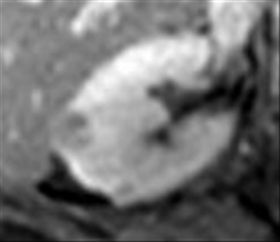
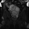
2 A 61-year-old female with an incidentally discovered renal mass undergoes an abdominal MRI. Which finding on T1-weighted dual-echo gradient-recalled echo (GRE) imaging at 1.5 Tesla is most indicative of a benign histology?
A. Homogeneous signal loss at a TE of 2.2 msec
B. Homogeneous signal loss at a TE of 4.4 msec
C. India ink artifact at the interface between the mass and the perinephric fat
D. India ink artifact at the interface between the mass and the kidney
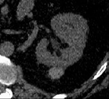
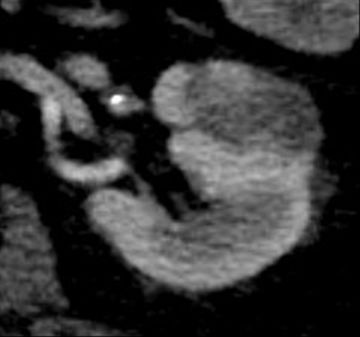
3 A 52-year-old male with autosomal dominant polycystic kidney disease presents with 12 hours of anuria and abdominal pain postoperative day 1 status post renal transplantation. Which of the following is the most likely explanation for the spectral waveform?
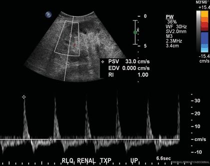
A. Renal artery stenosis
B. Renal arteriovenous fistula
C. Renal vein thrombosis
D. Normal superimposed waveforms
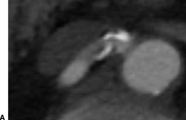
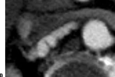
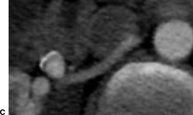
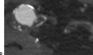
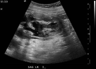
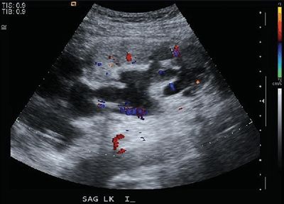
4 A 50-year-old male with hematuria and a renal transplant presents for Doppler imaging. What is the most likely explanation for the finding in the lower pole?
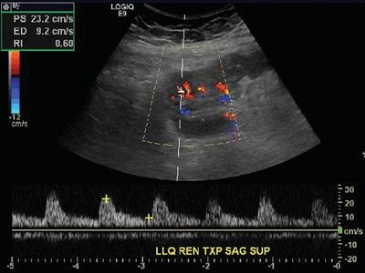
A. Polyarteritis nodosa
B. Iatrogenic injury
C. Congenital malformation
D. Renal artery thrombosis
E. Acute tubular necrosis
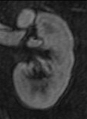
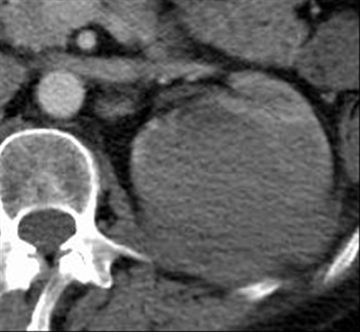
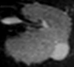
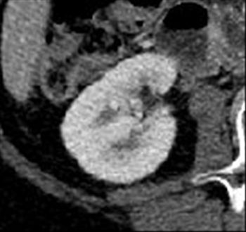
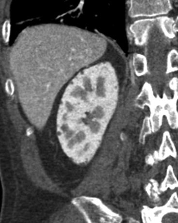
5 A 49-year-old male with an incidentally discovered renal mass undergoes a multiphasic renal mass protocol CT. This demonstrates a solitary exophytic 5.5-cm hypervascular heterogeneous solid right renal mass that lacks macroscopic fat and has a central scar. Which of the following is most correct?
A. The mass is a renal cell carcinoma.
B. The mass is an oncocytoma.
C. The mass is an angiomyolipoma.
D. The mass is a lymphoma.
E. The mass is a urothelial carcinoma.
F. The mass is indeterminate.
6 A 34-year-old female with numerous small solid echogenic renal masses undergoes a set of screening examinations. Which of the following studies is most likely to have been obtained in this patient?
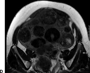
A.
B.
C.
D.
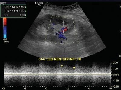
7 A 42-year-old male with diabetes mellitus type I complicated by end-stage renal disease undergoes a renal transplant. A percutaneous renal biopsy is performed 6 months later due to an elevated serum creatinine, and a subsequent renal ultrasound demonstrates an intrarenal arteriovenous fistula. What spectral Doppler waveform is characteristic of this entity?
A. High velocity, high resistance
B. High velocity, low resistance
C. Low velocity, high resistance
D. Low velocity, low resistance
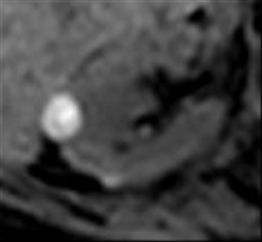
8 A 67-year-old male with a solid right renal mass undergoes percutaneous biopsy confirming clear cell renal cell carcinoma. Which artifact best explains the finding within the mass on T1-weighted dual-echo gradient-recalled echo (GRE) imaging at 1.5 Tesla?
Echo time (TE) = 2.2 msec
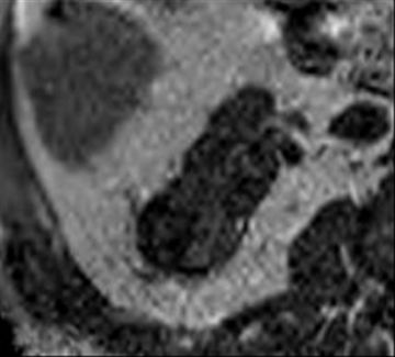
Echo time (TE) = 4.4 msec
A. Chemical shift artifact of the first kind
B. Chemical shift artifact of the second kind
C. Chemical shift artifact of the third kind
D. Chemical shift artifact of the fourth kind
9 A 60 year-old male with malaise and weight loss presents for contrast-enhanced CT, which demonstrates multiple bilateral non–contour-deforming low-attenuation renal masses (30 to 50 Hounsfield units). They are confirmed to be solid on MRI. Which of the following is the most likely diagnosis?
A. Tuberous sclerosis
B. Birt-Hogg-Dubé syndrome
C. Renal cell carcinoma
D. Urothelial carcinoma
E. Renal sarcoma
F. Lymphoma
G. von Hippel-Lindau syndrome
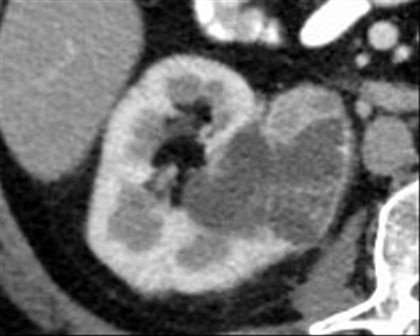
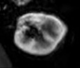
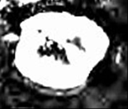
10 A 37-year-old female is found to have an incidental cystic renal mass. The highest attenuation component measures 180 Hounsfield units on postcontrast imaging (below) and 30 Hounsfield units on unenhanced imaging (not shown). What is the most appropriate Bosniak classification?
A. Bosniak I
B. Bosniak II
C. Bosniak IIF
D. Bosniak III
E. Bosniak IV
11 A 30-year-old female with chronic urolithiasis and multiple prior CT studies (one of which, performed 3 years ago, is shown below) presents with flank pain and hematuria. A renal stone CT is ordered. What is the best way to ensure that the radiation dose be as low as reasonably achievable (ALARA)?
A. Reduce the beam energy from 120 kVp to 80 kVp.
B. Reduce the tube current from 400 mA to 200 mA.
C. Enable tube current modulation.
D. Increase the pitch from 1.0 to 1.25.
E. Perform a renal ultrasound instead of a CT.
F. Enable adaptive statistical iterative reconstruction.
G. Apply breast and thyroid shielding.
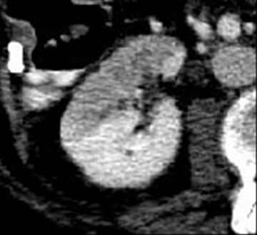
12 A 59-year-old female with hypertension treated with two oral medications has the following incidental finding. What is the best next step?
A. Partial nephrectomy
B. Percutaneous biopsy
C. Further imaging
D. Ignore. The finding is a normal variant.
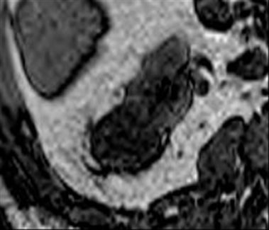
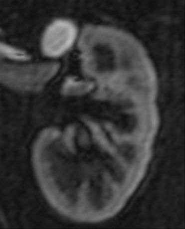
13 A 43-year-old female undergoes a multiphasic renal mass protocol CT of the abdomen demonstrating a 1.4-cm right renal mass. The attenuation measurements are 32 Hounsfield units on unenhanced images and 50 Hounsfield units on postcontrast images. An MRI is then obtained and is shown below. Given the MR findings, what is the best explanation for the measured attenuation difference on the unenhanced and postcontrast CT images?
T1-weighted precontrast
T1-weighted postcontrast
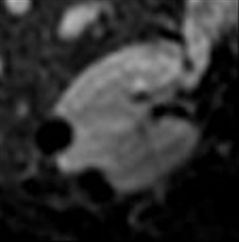
Subtraction
A. Solid enhancing tissue
B. Application of tube current modulation
C. Erroneous beam hardening correction
D. Respiratory motion artifact
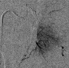
14 A 40-year-old female with recalcitrant hypertension undergoes CT angiography of the renal arteries. Which of the following would be treated best with balloon angioplasty alone?
A.
B.
C.
D.
15 A 59-year-old male with urosepsis and hypotension presents with 6 hours of absent urine output. A renal Doppler study is performed. Which of the following spectral Doppler waveforms is most characteristic of acute tubular necrosis?
A. Resistive index = 1.0 and minimal diastolic flow
B. Resistive index = 0.7 and intact diastolic flow
C. Resistive index = 0.4 and blunted systolic upstroke
D. Resistive index = 0.2 and elevated arterial velocity
16 A 62-year-old male with hematuria is found to have a large left renal mass. Which risk factor has the strongest association with the imaging findings?
A. Solid organ transplant
B. Obesity
C. Hypertension
D. von Hippel-Lindau syndrome

A. Anterior pararenal space
B. Posterior pararenal space
C. Perirenal space
D. Subcapsular space
18 A 45-year-old male was involved in a motor vehicle collision, and his left kidney was lacerated. Two weeks later, a contrast-enhanced CT was performed for persistent abdominal pain. What late-term complication is most strongly associated with the depicted abnormality?
A. Nephrolithiasis
B. Hypertension
C. Pyelonephritis
D. Fistula formation
19 A 55-year-old female undergoes an unenhanced CT of the abdomen demonstrating a renal mass that measures 62 Hounsfield units. There are no comparison studies. What is the best next step?
A. Observation
B. More imaging
C. Renal mass biopsy
D. Partial nephrectomy
20 A 2-year-old girl with abdominal swelling and pain undergoes a CT of the abdomen and pelvis. Which of the following organisms most likely colonizes this patient’s urinary tract?
A. Schistosoma haematobium
B. Proteus mirabilis
C. Mycobacterium tuberculosis
D. Staphylococcus aureus
E. Echinococcus multilocularis
21 A 52-year-old male presents with flank pain and dysuria, and a CT is performed 120 seconds after intravenous contrast material administration. Which of the following best describes the imaging finding in the left kidney?
A. Striated nephrogram
B. Delayed nephrogram
C. Absent nephrogram
D. Dense nephrogram
22 A 62-year-old male smoker with chronic obstructive pulmonary disease, coronary artery disease, hypertension, and peripheral vascular disease undergoes a CT angiogram of the abdomen and pelvis that demonstrates a 1.8-cm indeterminate homogeneous low-attenuation left renal mass (35 Hounsfield units). A renal mass protocol MRI is performed. What is the most likely explanation for the imaging findings?
T1-weighted precontrast
T1-weighted postcontrast
Subtraction
T2-weighted
T1-weighted precontrast (caudal to lesion)
Subtraction (caudal to lesion)
A. Chemical shift artifact
B. Misregistration artifact
C. Mural calcifications
D. Mural soft tissue
23 A 76-year-old patient with back pain undergoes a digital subtraction angiogram of the left kidney. What is the best next step?
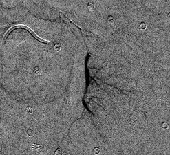
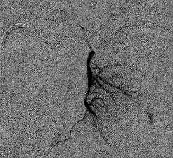
A. Covered stent
B. Balloon angioplasty
C. Embolization
D. Observation
E. Surgery
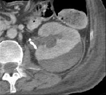
24 A 52-year-old male with biopsy-confirmed pancreatic adenocarcinoma undergoes a pancreaticoduodenectomy. During the postoperative course, a complication is suspected, and a contrast-enhanced CT of the abdomen is performed (shown below). Which of the following is the most specific imaging sign of acute renal infarction?
Preoperative study
Postoperative study (2 weeks later)
A. Alternating bands of hyper- and hypoattenuation extending to the capsule
B. Sharply demarcated cortical defect with an underlying calyceal diverticulum
C. Global delayed nephrogram with collecting system dilation and urothelial enhancement
D. Wedge-shaped focal delayed nephrogram with a thin enhancing capsular rim
25 A 51-year-old male with diabetes mellitus type II presents with right flank pain and dysuria. He has not seen a physician in many years. What is the best next step?
A. Operative debridement
B. Simple nephrectomy
C. Extracorporeal shock wave lithotripsy
D. Intravenous antibiotics
26 A 52-year-old female presents with acute right flank pain. A contrast-enhanced CT is performed. Which of the following risk factors is most likely to be present in this patient?
A. Relapsing urinary tract infection
B. Bladder cancer
C. Liver transplant recipient
D. Polycythemia vera
E. Recurrent nephrolithiasis
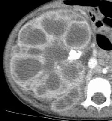
27 A 1-month-old infant presents with abnormal kidneys. Which of the following structures is likely to be also involved by this disease?
A. Spleen
B. Liver
C. Adrenal glands
D. Pancreas
E. Spinal cord
28 A 62-year-old male with diabetes mellitus type II, benign prostatic hypertrophy, and urinary retention presents with a 2-cm ill-defined left renal mass that is new since a comparison CT 6 months ago. Following a 2-week course of antibiotics, the imaging is repeated, demonstrating persistence of the finding. The mass enhances 30 Hounsfield units. What is the best next step?
A. Thermal ablation
B. Partial nephrectomy
C. Percutaneous biopsy
D. MRI
29 A 54-year-old male with no significant past medical history presents with fever, leukocytosis, tachycardia, hypotension, and dysuria. What is the definitive management for this condition?
A. Antibiotics
B. Percutaneous nephrostomy
C. Nephrectomy
D. Observation
30 A 60-year-old male presents for a contrast-enhanced CT of the abdomen, and an abnormality is noted in the kidneys. Which of the following is most strongly associated with this finding?
A. Hepatorenal syndrome
B. Mood disturbances
C. Untreated hypertension
D. Antihypoglycemic medications
Answers and Explanations
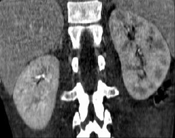
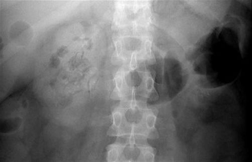
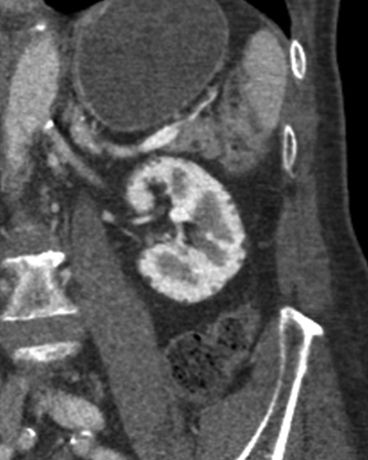
1 Answer B. The images demonstrate contrast material in the kidneys (corticomedullary phase), gallbladder (vicarious excretion), and bowel (oral contrast material), but no contrast material within the vasculature or other upper abdominal viscera. This suggests recent exposure to intravascular contrast material that is being abnormally retained within the kidneys (i.e., bilateral delayed nephrograms). The most common antecedent history in this context is a recent contrast-enhanced CT or coronary angiogram in the setting of hypotension. The generic differential diagnosis for a delayed nephrogram can be oversimplified as follows:
- “Blood in”: renal artery stenosis/thrombosis/laceration
- “Blood out”: renal vein thrombosis
- “Urine in”: acute tubular necrosis, pyelonephritis, glomerulonephritis
- “Urine out”: collecting system obstruction
In this case, the delayed nephrogram is diffuse and bilateral. Therefore, acute tubular necrosis is the best answer. Contrast-induced nephrotoxicity (Answer A) could cause this appearance, but it is not the best answer because it is an uncommon and specific cause of acute kidney injury. Although pyelonephritis can be bilateral (Answer C), it would more likely present with striated nephrograms. In this case, the pattern of enhancement is globally delayed, not striated. Although extracellular gadolinium-based contrast material can resemble dilute iodinated contrast material on CT (Answer D), it in general follows the same dynamic enhancement pattern. In this case, the enhancement pattern is abnormal; therefore, gadolinium-based contrast material exposure is not the best answer.
References: Dyer RB, Munitz HA, Bechtold R, et al. The abnormal nephrogram. Radiographics 1986;6:1039–1063.
Saunders HS, Dyer RB, Shifrin RY, et al. The CT nephrogram: implications for evaluation of urinary tract disease. Radiographics 1995;15:1069–1085.
Wolin EA, Hartman DS, Olson JR. Nephrographic and pyelographic analysis of CT urography: differential diagnosis. AJR Am J Roentgenol 2013;200:1197–1203.
Related posts:
Stay updated, free articles. Join our Telegram channel

Full access? Get Clinical Tree


