A. Shattered kidney, operative management
B. Renal contusions, conservative management
C. Pyelonephritis, antibiotic therapy
D. Renal infarcts, angiography and/or echocardiography
2 A retrospective cohort study evaluating the incidence of contrast-induced acute kidney injury (CI-AKI) compares acute kidney injury rates following CT in two groups: (1) patients who received contrast-enhanced CT as part of routine clinical care and (2) patients who received unenhanced CT as part of routine clinical care. Serum creatinine measurements drawn 48 hours after the CT are compared to serum creatinine measurements drawn prior to the CT. An increase at the 48-hour time point of ≥0.3 mg/dL is considered indicative of CI-AKI. All patients included in the study had all necessary serum creatinine measurements available. What source of bias is of greatest concern with this type of study design?
A. Verification bias
B. Selection bias
C. Recall bias
D. Lead-time bias
3 A 70-year-old male with a dilated left collecting system undergoes a retrograde pyelogram. What entity is classically associated with the imaging findings?
A. Urothelial carcinoma
B. Calculus disease
C. Blood clot
D. Fungal infection
E. Schistosoma haematobium
4 A 52-year-old female with chronic obstructive pulmonary disease on 2 L of home oxygen therapy undergoes a CT for right lower quadrant pain. A 1.5-cm cortically based left renal mass is identified. Biphasic renal mass protocol CT is performed demonstrating the following features: (1) unenhanced attenuation 50 HU, (2) nephrographic phase attenuation 100 HU, (3) no macroscopic fat, and (4) homogeneous enhancement. The unenhanced background renal parenchyma is 30 HU, the adjacent renal artery is 180 HU, and there are no comparison studies. What is the best next step?
A. Ignore (benign finding)
B. Percutaneous biopsy
C. Partial nephrectomy
D. Embolization
5 A new blood test is developed for prostate cancer screening. When the test is positive, a transrectal ultrasound (TRUS)-guided random prostate biopsy is performed. After decades of study, it is determined that patients diagnosed with prostate cancer through the screening program have improved survival. However, it is repeatedly noted that prostate cancers resulting from a screening-triggered biopsy are more likely to be variants with a slower growth rate. What type of screening bias results from the disproportionate identification of slowly progressive disease?
A. Reporting bias
B. Lead-time bias
C. Length bias
D. Overdiagnosis bias
6 Calculate the approximate voxel size of the 2D T2-weighted fast spin echo image shown below provided the following assumptions: (1) field of view = 16 cm × 16 cm, (2) phase-encoding matrix = 168, (3) frequency-encoding matrix = 228, (4) TE = 110 msec, (5) TR = 5,190 msec, (6) slice thickness = 3 mm, (7) interslice gap = 1 mm, (8) echo train length = 21.
A. 0.95 mm × 0.70 mm × 3.00 mm
B. 0.95 mm × 0.70 mm × 4.00 mm
C. 1.43 mm × 1.05 mm × 3.00 mm
D. 1.43 mm × 1.05 mm × 4.00 mm
E. 0.14 mm × 0.11 mm × 3.00 mm
F. 0.10 mm × 0.07 mm × 4.00 mm
G. The voxel size cannot be calculated with the provided information.
7 A 59-year-old female with hematuria undergoes an intravenous pyelogram (IVP) demonstrating a mass in the left renal pelvis. What is the main reason that CT urography (CTU) has supplanted IVP for the detection of upper tract malignancy?
A. CTU requires less radiation than IVP.
B. CTU uses a lower dose of contrast material than IVP.
C. CTU detects less incidental findings than IVU.
D. CTU has a higher accuracy than IVU.
8 A 55-year-old female with no comorbidities undergoes percutaneous core biopsy of a 3.2-cm solid mass in the right perinephric space. The histology demonstrates epithelioid angiomyolipoma. What is the best next step?
A. Observation (benign finding)
B. Repeat biopsy
C. Embolization
D. Operative resection
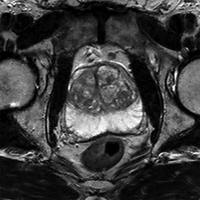
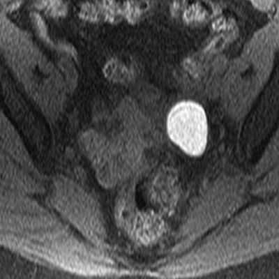
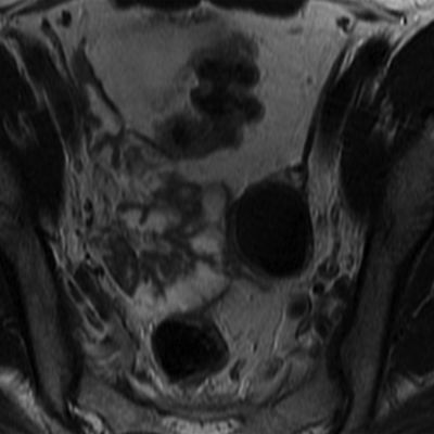
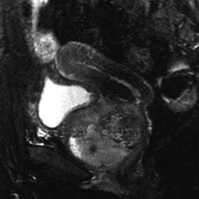
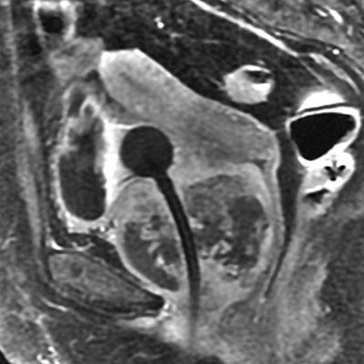
9 A 75-year-old male with elevated prostate specific antigen (PSA, 25 ng/mL) undergoes a 3.0-Tesla prostate MRI. What is the hyperintense elliptical structure identified along the midline?
A. Glandular BPH nodule
B. Stromal BPH nodule
C. Utricle cyst
D. Prostate cancer
10 A 73-year-old male with Gleason 4 + 3 = 7 prostate cancer undergoes a 3.0-Tesla prostate MRI. What can be done to reduce the artifacts within the prostate gland?
A. Change the frequency-encoding axis to: “left-to-right”
B. Change the phase-encoding axis to: “left-to-right”
C. Apply fat saturation using short tau inversion recovery (STIR)
D. Apply fat saturation using a spectrally selective radiofrequency pulse
11 A 50-year-old female with a left renal angiomyolipoma presents for treatment. At what size is embolization commonly considered for the treatment of an asymptomatic angiomyolipoma?
A. ≥2 cm
B. ≥4 cm
C. ≥6 cm
D. ≥8 cm
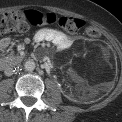
12 A 30-year-old male with acute left flank pain undergoes an unenhanced CT of the abdomen and pelvis. What is the most likely explanation for the abnormality in the left perinephric space?
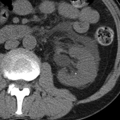
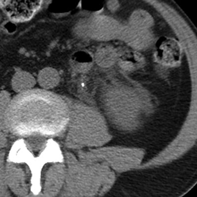
A. Urinoma
B. Abscess
C. Edema
D. Lymphoma
13 A 75-year-old female with stage IV chronic kidney disease (estimated glomerular filtration rate is 19 mL/min/1.73 m2), hypertension, peripheral vascular disease, and claudication symptoms presents for a CT angiogram of the abdomen and pelvis with runoff to the lower extremities. Which of the following prophylactic measures has the most supportive evidence as a technique to reduce this patient’s risk of contrast-induced acute kidney injury?
A. Volume expansion with IV D5 0.45% NaCl
B. Volume expansion with IV 0.9% NaCl
C. Preprocedure administration of N-acetylcysteine
D. Preprocedure administration of sodium bicarbonate
14 A 70-year-old male presents for an ultrasound-guided percutaneous biopsy of an incidentally detected 2-cm homogeneously enhancing solid renal mass. What is the most common complication of this procedure?
A. Hemorrhage
B. Needle-track seeding
C. Renal abscess
D. Pneumothorax
E. Collecting system obstruction
F. Colonic injury
15 A 70-year-old female with a 2.0-cm heterogeneous solid renal mass presents for percutaneous renal mass biopsy. Which of the following approaches is most likely to yield a diagnostic result with the least risk of complication using an 18-gauge spring-loaded end-firing core biopsy device with a 1.0-cm or 2.0-cm throw length?
A. Angle A with a 1-cm throw length
B. Angle A with a 2-cm throw length
C. Angle B with a 1-cm throw length
D. Angle B with a 2-cm throw length
E. Angle C with a 1-cm throw length
F. Angle C with a 2-cm throw length
16 A 50-year-old female with back and flank pain undergoes an abdominal MRI. What is the most likely cause of the signal abnormality anterior to the common iliac arteries?
A. Magnetic field inhomogeneity
B. Inaccurate inversion time
C. Retroperitoneal fibrosis
D. Lymphoma
17 A 63-year-old male with metastatic prostate cancer undergoes a CT of the abdomen and pelvis. How should the current images be interpreted?
Current study Nine months ago
A. Osseous metastatic disease; uncertain response
B. Osseous metastatic disease; progressive disease
C. Osseous metastatic disease; partial response
D. Osseous metastatic disease; stable disease
18 A 63-year-old male presents with scrotal discomfort and an ultrasound is performed. What is the most likely diagnosis?
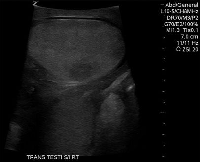
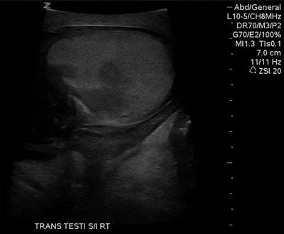
A. Seminomatous germ cell tumor
B. Nonseminomatous germ cell tumor
C. T-cell lymphoma
D. B-cell lymphoma
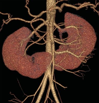
A. Crossed-fused ectopia
B. Horseshoe kidney
C. Pelvic kidney
D. Duplicated kidney
20 A 28-year-old female presents with lower abdominal pain and a CT is performed. What is the most likely antecedent history?
A. Family history of ovarian cancer
B. Hyperandrogenism and metabolic syndrome
C. Recent normal menstruation
D. Recent fertility treatment
21 A 40-year-old female with vague abdominal pain undergoes a CT of the abdomen and pelvis. What is the most likely diagnosis?
A. Lymphoma
B. Liposarcoma
C. Angiomyolipoma
D. Vascular malformation
E. Foreign body
22 A 33-year-old female with left lower quadrant pain undergoes a pelvic MRI. A nonenhancing abnormality is identified within the left ovary. What is the most likely cause of the T1 shortening within this abnormality?
A. Fat
B. Protein
C. Hemorrhage
D. Melanin
E. Contrast material
F. Flow artifact
G. Calcium
23 A 50-year-old female with pelvic pain undergoes a pelvic MRI and a large necrotic mass is identified. What is the most likely diagnosis?
A. Cervical cancer
B. Vaginal cancer
C. Urethral cancer
D. Bladder cancer
E. Prostate cancer
24 A 70-year-old male with an incidentally detected 2-cm solid renal mass presents for a discussion regarding management. What is the commonly cited size threshold at which renal cell carcinoma is prone to metastasize?
A. 1 cm
B. 3 cm
C. 5 cm
D. 7 cm
25 Which of the following patients has the greatest risk of nephrogenic systemic fibrosis upon administration of gadolinium-based contrast material?
A. A 60-year-old male with acute kidney injury and eGFR 55 mL/min/1.73 m2
B. A 40-year-old female with diabetes mellitus and eGFR 40 mL/min/1.73 m2
C. A 50-year-old male with severe hypertension and eGFR 45 mL/min/1.73 m2
D. A 70-year-old female with multiple myeloma and eGFR 60 mL/min/1.73 m2
26 A radiology practice is interested in reducing radiation exposure to patients undergoing CT urography. Assuming that each of the following preserves the diagnostic accuracy of the examination, which will have the greatest effect on radiation dose reduction?
A. A 20% decrease in mA
B. A 20% decrease in kVp
C. A 20% decrease in pitch
D. A 20% decrease in z-axis coverage
27 A 70-year-old female with peripheral vascular disease and a suspected paraneoplastic syndrome undergoes a CT of the chest, abdomen, and pelvis. A 1.2-cm ovoid retrocrural mass is identified that measures 12 Hounsfield Units. How should this finding be interpreted?
A. Normal variant, ignore
B. Enlarged lymph node, recommend biopsy
C. Enlarged lymph node, recommend upper endoscopy
D. Suspected paraganglioma, recommend MIBG
E. Suspected benign nerve sheath tumor, ignore
28 A 45-year-old average-size male with a psoas abscess is given conscious sedation with midazolam IV and fentanyl IV for an ultrasound-guided drainage catheter placement. During the procedure, the patient complains of ongoing back pain, and so three additional doses of fentanyl IV are administered at 5, 10, and 15 minutes. At 20 minutes, the patient becomes hypoxic and his respiratory rate is 6. Verbal and tactile stimulation has minimal effect. What is the best next step?
A. Administer naloxone 0.2 mg IV q2–3 min until his respiratory rate is >10
B. Administer naloxone 2.0 mg IV q2–3 min until his respiratory rate is >10
C. Administer flumazenil 0.1 mg IV, may repeat q1 min, goal is respiratory rate >10
D. Administer flumazenil 1.0 mg IV, may repeat q1 min, goal is respiratory rate >10
29 A 55-year-old male with new-onset proteinuria is being scheduled for an elective renal biopsy. He has a history of atrial fibrillation and is receiving chronic warfarin therapy. According to the Society of Interventional Radiology 2012 consensus guidelines, what should be the target INR (International Normalized Ratio) for this procedure?
A. INR < 1.9
B. INR < 1.7
C. INR < 1.5
D. INR < 1.3
30 A 20-year-old male with upper abdominal pain undergoes a contrast-enhanced CT of the abdomen and pelvis, and an abnormality is identified in his pelvis. Which of the following is strongly associated with this imaging finding?
A. Chronic urinary retention
B. Ipsilateral renal agenesis
C. Chlamydia trachomatis infection
D. Prostate cancer
Answers and Explanations
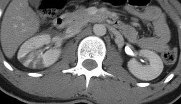
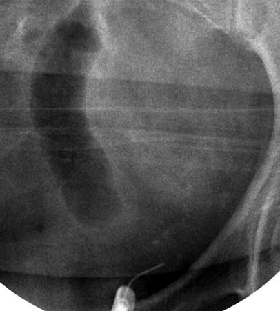
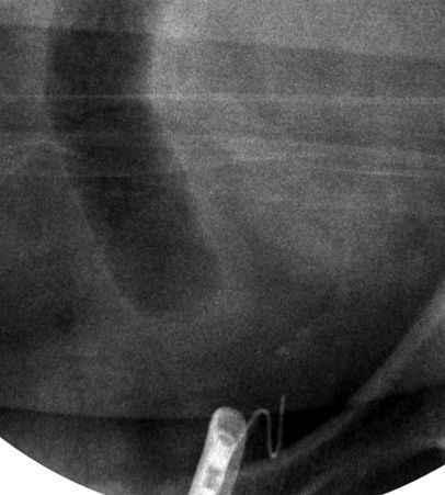
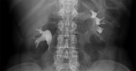
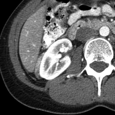
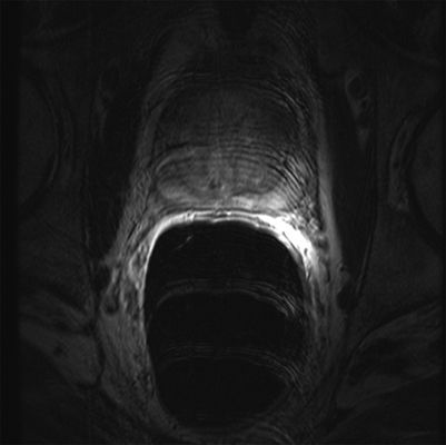
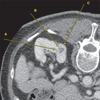
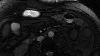
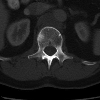
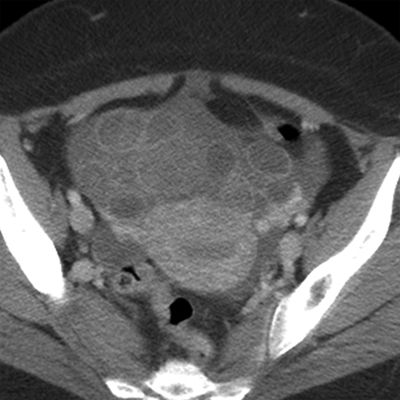
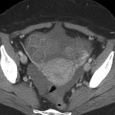
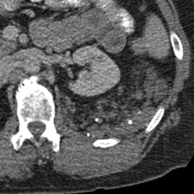
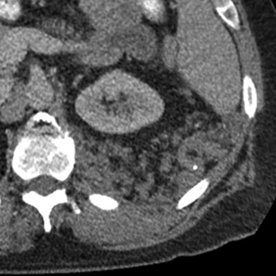
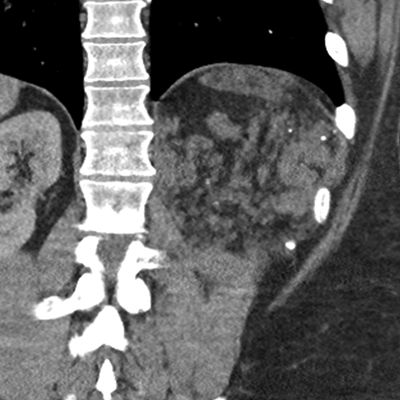
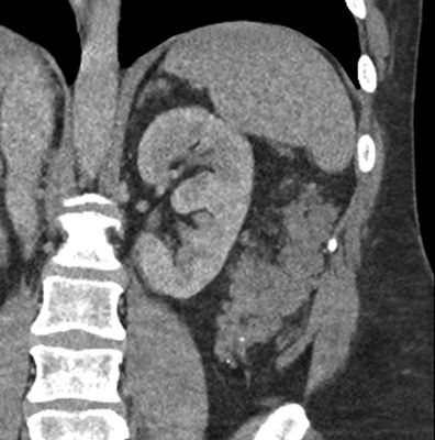
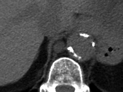
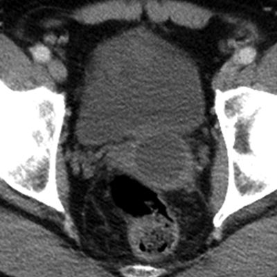
1 Answer B. The images demonstrate a striated right nephrogram in the setting of trauma indicating multifocal contusion and/or laceration. This should be managed conservatively because the vessels are uninjured, the hilum and collecting system are intact, and there is only a small hematoma in the perinephric space. Follow-up imaging performed 2 years later (below) reveals cortical scarring at each site of injury but an otherwise intact, functioning kidney.
Other causes of a striated nephrogram include: (1) infarcts from a peripheral source (e.g., polyarteritis nodosa), (2) infarcts from a central source (e.g., endocarditis, renal artery injury), (3) renal vein thrombosis, (4) pyelonephritis, (5) glomerulonephritis, and (6) urinary tract obstruction.
A striated nephrogram is basically a multifocal incomplete delayed nephrogram in which some parts of the kidney are affected more than others. The differential diagnosis for a striated nephrogram is therefore similar to the differential diagnosis for a delayed nephrogram and follows a very simple rubric: (1) blood in, (2) blood out, (3) urine in, (4) urine out; when one or more of these four processes is disrupted, a delayed (or striated) nephrogram can result.
References: Kawashima A, Sandler CM, Corl FM, et al. Imaging of renal trauma: a comprehensive review. Radiographics 2001;21:557–574.
Santucci RA, McAninch JW, Safir M, et al. Validation of the American Association for the Surgery of Trauma organ injury severity scale for the kidney. J Trauma 2001;50:195–200.
Saunders HS, Dyer RB, Shifrin RY, et al. The CT nephrogram: implications for evaluation of urinary tract disease. Radiographics 1995;15:1069–1085.
Related posts:


Stay updated, free articles. Join our Telegram channel

Full access? Get Clinical Tree


