Fig. 11.1
A young girl that fell off her horse. Plain film (a), CT (b), and MR with sagittal T1–WI (c), T2–WI (d), and STIR (e). This 17-year-old girl suffered an unusual spinal lesion for a horse riding accident. Although cervical lesions form a minority of spinal fractures (<10 %) in this sport, they are the most feared lesions because of the possibility of permanent neurological dysfunction and even death. Plain films showed a wedge compression fracture of C4, and subsequent CT revealed a sagittal and a coronal fracture through the vertebral body making this an instable lesion. Because of paresthesias and motor weakness in both arms, a neuropraxia of the cervical spinal cord was diagnosed. Although there was a rapid and complete resolution of symptoms, MRI was warranted. No medullary lesion was seen. STIR images reveal bone bruises to C7 and Th2 and lesions of the intraspinous ligaments at C4–C5, C5–C6, C6–C7, and Th1–Th2 in this hyperflexion trauma

Fig. 11.2
A 6-year-old boy that dived off a ledge in shallow water. Lateral (a) and open-mouth (b) plain film. When diving in shallow water and landing on the head with axial loading of the spine, typically cervical spine lesions are found. The most typical fracture is of the ring of C1, a so-called Jefferson fracture. It was originally described as a 4-part fracture with double fractures through the anterior and posterior arches, but 3-part and 2-part fractures have also been described. It results from the occipital condyles being driven into the lateral masses of C1. This case recovered completely without surgical intervention
Fig. 11.3
A 19-year-old with cervical pain after a rugby game. Plain film (a), CT (b, c). This young man was sitting in the X-ray department waiting room on Monday morning with persisting cervical pain after a rugby game on Sunday. AP plain film shows a lateral C6–C7 dislocation. CT confirms the dislocation and demonstrates a unilateral facet fracture. Always carefully examine the soft tissue windows (c) also: in this case, a large epidural hematoma is shown while compressing the dural sac
Fig. 11.4
Low thoracic burst fracture: lateral film (a), axial CT (b). Th12 burst fracture in a young woman that fell off her horse. Th12, L1, and L2 burst fractures are the most common spinal lesions in horseback riding accidents. They occur when riders fall of, or are thrown off, of their horse and land on their buttocks or lower back. Although plain films are helpful in the diagnosis, CT is obligatory in order to evaluate lesion stability and displacement of bone in the spinal canal and/or foramina
11.2.1.2 Muscle and Ligamentous Lesions
Sprain represents stretching of a ligament beyond its elastic limit. Diagnostic imaging is negative except for ultrasonography or MRI that may show edema in the soft tissues, muscle tear, and/or hematoma (Fig. 11.5). Imaging is not necessary if there is no neurologic deficit and if the injury did not occur with an excessive amount of force or if fracture is to be excluded (Haus 2012; Trainor 2002).
Fig. 11.5
Low back soft tissue lesion in a trampoline accident: MR with sagittal T1–WI (a), T2–WI (b), and STIR (c). This 14-year-old girl suffered a trampoline accident with acute low back pain. Plain films and CT were normal. MRI was performed several days later because of persisting severe low back pain. T1–WI and T2–WI show subtle signal intensity changes in the upper/anterior part of the vertebral bodies L2, L3, and L4. These bone bruises are much more obvious on the STIR images confirming the need for robust fat-suppressed imaging. Moreover, there are lesions of the intraspinous ligaments L1–L2 and especially L2–L3 confirming a flexion-type injury
11.2.1.3 Disc Injuries
Discogenic pathology, including disc herniation, is responsible for 11 % of lumbar pain in pediatric athletes. The majority of athletes with a disc herniation are older than the age of 12. The most common locations for disc herniations are at the L4–L5 and L5–S1 levels (92 % of cases in adolescents). Most of the disc herniations remain subligamentous. Athletes at risk are those participating in collision sports and weight lifting due to the increased axial forces exerted on the spine. Often these patients have underlying vertebral anomalies such as scoliosis, transitional vertebrae (lumbarization and sacralization), lysis or spinal canal narrowing that may cause early disc degeneration, and subsequent herniation. Pain is typically located in the low back with or without leg pain. Overt neurologic deficits are rare. MRI remains the best modality to assess the herniated disc (Haus 2012).
11.2.1.4 Spinal Cord and Nerve Root Injuries
Cervical spinal cord injuries have been the most common catastrophic football injury and the second leading direct cause of death attributable in American football (Cantu and Mueller 2003). The relatively common “stinger” is a neuropraxia of a cervical nerve root(s) or brachial plexus and represents a reversible peripheral nerve injury. Less common and more serious an injury, cervical cord neuropraxia is the clinical manifestation of neuropraxia of the cervical spinal cord due to hyperextension, hyperflexion, or axial loading (Rihn et al. 2009).
11.2.2 Chronic Overuse Spinal Injuries
Spinal injuries in chronic overuse occur due to repetitive training and microtrauma. In children and adolescents, they are a major source of morbidity and in fact exceed infectious diseases as a cause of morbidity (Haus 2012). The prevalence of specific etiologies of back pain is different depending on the age of the athletes. The most frequent source of back pain in adolescents is spondylolysis; in young adults, disc disease is most frequent, and in older subjects, facet joint disease is most prevalent (Micheli 1995).
Recently, the more competitive nature of organized sports and the increased training load, necessary to obtain higher levels of competence, result in more repetitive microtrauma that may lead to overuse failure of spinal structures.
11.2.2.1 Spondylolysis
Radiography
When spondylolysis is suspected, anteroposterior, lateral, oblique left and right, and collimated lateral views of the spine are performed. Using these five views, most spondylolytic defects are detected. In the acute setting, the defect is narrow with irregular borders, whereas more chronic lesions have smoother and more rounded edges. The width of the gap depends on the grade of spondylolisthesis. Indirect signs of spondylolysis on the AP view are lateral deviation of the spinous process and sclerosis of the contralateral pedicle. The oblique views may show the “scotty dog” sign (a break in the neck of the dog representing the isthmic defect). Although 45° oblique views have long been thought to be the best projections for detecting spondylolysis, the study is only 32 % sensitive (Haus 2012). They demonstrate isthmic defects only well if the X-ray beam is perpendicular to the pars, while the orientation of lysis is in the majority of cases close to the coronal plane. This is why the collimated lateral view actually shows the majority of defects (Leone et al. 2011). Also a hypoplastic L5 vertebral body is evidence of an L5 spondylolytic defect. The forward displacement in spondylolisthesis is determined on the lateral view. Listhesis can be graded according to the percentage of slipping by dividing the subjacent vertebral body into four equal parts. A slip in the first quarter is grade I and a slip in the last quarter is grade IV. In symptomatic spondylolisthesis, dynamic flexion and extension views should be obtained to determine lumbar intervertebral instability. Most spondylolisthesis occur in flexion. Axial or extension spondylolisthesis more often give nerve root compression. Sometimes this is the only anomaly found in young people with low back pain complaints. The distinction between normal and abnormal movement is however difficult with a substantial overlap between symptomatic and asymptomatic motion patterns. General values of 10 % for sagittal rotation (i.e., variation of the angle between vertebral endplates adjacent to the disc) and 4 mm for sagittal translation (i.e., slip of one vertebra relative to the vertebra below) are used as cutoff.
Single-Photon Emission Computed Tomography
When radiographs are negative for spondylolysis, but clinical suspicion is high, the more sensitive test is single-photon emission computed tomography (SPECT). SPECT is useful in early stages as it can identify patients with symptomatic (pre-)spondylolysis before radiography does. Positive SPECT suggests symptomatic spondylolysis, stress reaction, stress fracture, or healing (Fig. 11.6). It can differentiate between acute spondylolysis and a nonunion of the pars. Radiographic pars defects without uptake on SPECT are often asymptomatic. There has, however, also been documented positive SPECT imaging in asymptomatic athletes. Furthermore, activity on SPECT is nonspecific and has to be differentiated from other entities, for example, facet arthritis, infection, or osteoid osteoma. Therefore, if SPECT is positive, a CT scan should be performed for further evaluation. Therefore, SPECT is not recommended as screening tool in young patients. SPECT can be used to guide therapy. SPECT-positive spondylolytic lesions may better respond to conservative therapy (Leone et al. 2011; Papanicolaou et al. 1985; Maxfield 2010).
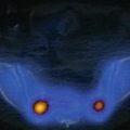
Fig. 11.6


Young gymnast with subacute/chronic low back pain: axial CT (a), parasagittal CT right (b) and left (c) (CT performed as part of a SPECT-CT exam), parasagittal STIR MR right (d) and left, (e) and SPECT-CT (f). These are images of a 14-year-old gymnast. Because of persisting low back pain, imaging was performed. When suspecting spondylolysis, CT is a very sensitive and specific imaging modality. In this case, a bilateral spondylolysis is shown although it may be incomplete on the right. MRI confirms an active process with edematous changes bilaterally but more extensive edema on the left. On the right (d), there is a band of low signal intensity at the spondylolytic lesion. SPECT-CT is asymmetric showing more activity on the right. The reduced bone edema with more SPECT activity on the right might be an indication of healing
Related posts:


Stay updated, free articles. Join our Telegram channel
Full access? Get Clinical Tree
