Chapter 14 Sacrum and coccyx
Sacrum
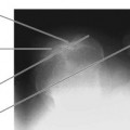
The sacrum may be a site for metastatic spread of malignancy and plain images of the region would demonstrate such lesions if the secondary tumour has eroded at least 40% of the bone. Magnetic resonance imaging (MRI) is most appropriate for assessing bone metastases.1 Today it is rare to find radiography requested for assessment of this area, and there is no longer specific reference to assessment of the sacrum in current Royal College of Radiologists (RCR) guidelines in the UK.
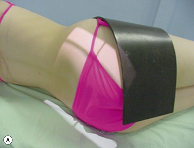
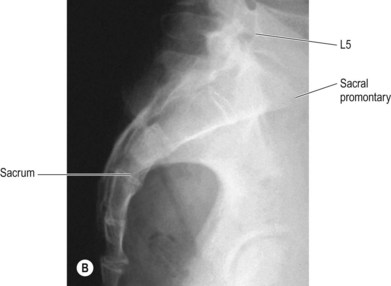
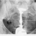
Lateral sacrum (Fig. 14.1A,B)
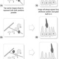
Positioning
• The patient lies on their side, with hips and knees flexed to maintain stability and the feet placed together to prevent the patient from rolling forwards or backwards. The arms are flexed at the elbow and raised to rest on the pillows for comfort and to clear them from the area of interest
• Lead rubber is applied over the raised side, diagonally from anterior superior iliac spine (ASIS) to the femoral head, to cover the anterior portion of the pelvis and to protect the gonads
• The palm of the radiographer’s hand is used to palpate the posterior aspect of the sacrum and ensure that its transverse axis is perpendicular to the table-top
• The long axis of the sacrum is parallel to the table-top; this should be checked with the area at the radiographer’s eye level for accuracy. If patient build affects the relationship of the sacrum to the table-top, a compensating cranial or caudal central ray can be used (see beam direction, below)
Beam direction and focus receptor distance (FRD)
Vertical, directed at 90° to the long axis of the sacrum once this has been assessed
Collimation
Lumbosacral junction, sacral promontory, soft tissues overlying the sacrum posteriorly, coccyx
Criteria for assessing image quality
• Lumbosacral junction, sacral promontory, soft tissues overlying the sacrum posteriorly and coccyx are demonstrated. Omission of the coccyx from the field may be acceptable if demonstration of the coccyx is not required specifically for the examination
• Joint space at the lumbosacral junction is demonstrated
• Sharp image demonstrating bony trabeculae. Adequate penetration to demonstrate detail of sacrum and less dense coccyx on the one image

Stay updated, free articles. Join our Telegram channel

Full access? Get Clinical Tree


