I Liver
CASE 1
Clinical Presentation
A 54-year-old man presents with nonspecific abdominal discomfort.

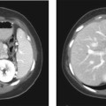
Fig. 1.1 Hepatic hemangioma. (A) Contrast-enhanced axial CT image of the liver shows a well-defined hypodense lesion with peripheral, nodular contrast enhancement (arrows) during the portal phase. The density of the nodular enhancement is similar to the density of the opacified aorta. (B) Delayed contrast-enhanced axial CT image of the liver shows irregular fill-in of contrast within the lesion.
Radiologic Findings
Contrast-enhanced computed tomography (CT) scan images of the liver show a large hypodense lesion with peripheral nodular enhancement with density of nodules similar to the density of the opacified aorta (Fig. 1.1A); there is centripetal enhancement and retention of contrast on the more delayed series (Fig. 1.1B).
Diagnosis
Hepatic hemangioma
Differential Diagnosis
- Liver metastases
Discussion
Background
Hemangioma is the most common tumor of the liver. The incidence of hemangioma in the general population is reported to be ˜ 20%.
Clinical Findings
Typically, hemangiomas are found in middle-aged women and are usually asymptomatic. When large they can cause symptoms secondary to complications.
Complications
Large hemangiomas can undergo spontaneous or traumatic rupture. There can be spontaneous bleeding within these lesions. Large hemangiomas can exert mass effect on adjacent structures, resulting in obstruction of hepatic vessels or the biliary tree, or can lead to Kasabach-Merritt syndrome, which is a form of consumptive coagulopathy due to thrombocytopenia. High-output congestive cardiac failure has also been reported related to large lesions. Pedunculated hemangiomas, regardless of size, can torse, leading to thrombosis and necrosis.
Etiology
Related posts:


Stay updated, free articles. Join our Telegram channel

Full access? Get Clinical Tree


