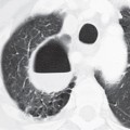
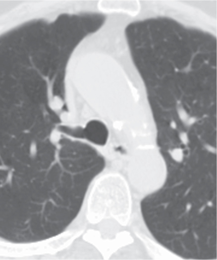
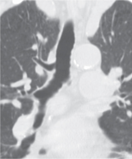
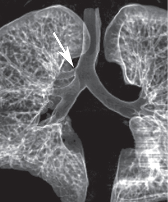
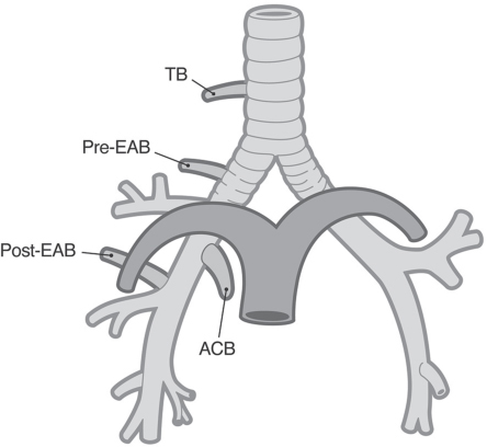
CASE 1 74-year-old man evaluated for exacerbation of chronic obstructive pulmonary disease Axial and coronal chest CT (lung window) (Figs. 1.1, 1.2) demonstrates a right tracheal bronchus. A normal right upper lobe apical segmental bronchus was not present. Volume-rendered surface display (Fig. 1.3) demonstrates an anomalous bronchus arising from the right lateral tracheal wall (arrow). Artist’s illustration (Fig. 1.4) depicts variations of right-sided anomalous bronchial branching. The tracheal bronchus (TB) arises from the lateral tracheal wall. The pre-eparterial bronchus (Pre-EAB) arises from the right mainstem bronchus proximal to the origin of the right upper lobe bronchus. The post-eparterial bronchus (Post-EAB) arises distal to the origin of the right upper lobe bronchus. The accessory cardiac bronchus (ACB) arises from the medial wall of the right mainstem bronchus or the bronchus intermedius and is usually blind-ending. Tracheal Bronchus • Tracheal Diverticulum • Tracheal Air Cyst Fig. 1.1 Fig. 1.2 Fig. 1.3 Fig. 1.4 Variations of bronchial branching are common, may affect all portions of the tracheobronchial tree, and are usually discovered incidentally. Tracheal bronchus is characterized by its anomalous origin from the lateral tracheal wall (usually within 2.0 cm from the carina) and typically supplies the right upper lobe. It is also known as “pig bronchus” or “bronchus suis” (the usual anatomic bronchial morphology in pigs and certain other mammals). Other right-sided anomalous bronchi are classified as pre-eparterial or post-eparterial
 Clinical Presentation
 Radiologic Findings
 Diagnosis
 Differential Diagnosis
 Discussion
Background
![]()
Stay updated, free articles. Join our Telegram channel

Full access? Get Clinical Tree


Radiology Key
Fastest Radiology Insight Engine

