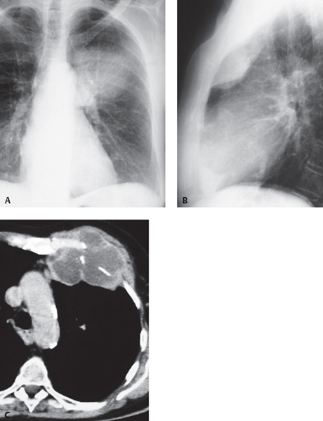
CASE 182 46-year-old man with chest pain and a palpable left anterior chest wall mass Coned-down PA (Fig. 182.1A) and lateral (Fig. 182.1B) chest radiographs demonstrate a left anterior chest wall mass. The lesion exhibits a well-defined lobulated border with obtuse angles against the adjacent chest wall on the lateral radiograph (Fig. 182.1B) and manifests as a left mid-thoracic opacity with poorly defined margins on the frontal radiograph (Fig. 182.1A). Unenhanced chest CT (mediastinal window) (Fig. 182.1C) shows a heterogeneous left anterior chest wall mass arising at the costochondral-sternal junction with multi-focal dense, linear calcifications. Fig. 182.1 Chondrosarcoma • Metastasis from Primary Chondrosarcoma or Other Primary Malignant Neoplasm • Primitive Malignant Neuroectodermal Tumor • Malignant Fibrous Histiocytoma • Fibrosarcoma; Neurofibrosarcoma Chondrosarcomas are malignant neoplasms with cartilaginous differentiation. They represent the most common primary malignant tumor of the chest wall. They typically arise in the anterior chest wall and involve the sternum or costochondral arches. Less frequently they arise in the ribs (17%) and scapulae. Chondrosarcomas occur across a wide age range but typically affect patients between the ages of 30 and 60 years. Most tumors manifest as a palpable chest wall mass that may be painful. The lesion may grow rapidly and cause chest pain. Males are affected slightly more frequently than females (male to female ratio of 1.3:1.0).
 Clinical Presentation
 Radiologic Findings
 Diagnosis
 Differential Diagnosis
 Discussion
Background
Clinical Findings
Imaging Findings
Chest Radiography
Related posts:


Stay updated, free articles. Join our Telegram channel

Full access? Get Clinical Tree


