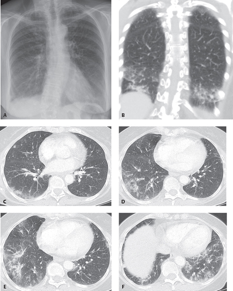
CASE 105 46-year-old woman, non-smoker, complaining of dyspnea and cough over the past six to eight months PA (Fig. 105.1A) chest radiograph demonstrates subtle bilateral ground glass and reticular opacities most pronounced in the mid- and lower lung zones. Coronal (Fig. 105.1B) and axial (Figs. 105.1C, 105.1D, 105.1E, 105.1F) CT (lung window) confirms the presence of primarily lower lobe patchy ground glass opacities and mild reticulation without significant fibrosis. Note the relative juxtapleural sparing by ground glass in the dorsal regions of the lower lobes. Open lung biopsy confirmed the diagnosis. Non-Specific Interstitial Pneumonia (NSIP); Cellular Pattern • Acute Interstitial Pneumonia (AIP) • Cryptogenic Organizing Pneumonia (COP) • Desquamative Interstitial Pneumonia (DIP) • Hypersensitivity Pneumonitis (HP) • Respiratory Bronchiolitis–Interstitial Lung Disease (RB-ILD) • Usual Interstitial Pneumonia (UIP) Non-specific interstitial pneumonia (NSIP) is one of the chronic idiopathic interstitial pneumonias (IIPs). Katzenstein first described NSIP in 1994. The term is used for cases of interstitial pneumonia in which diagnostic features of UIP, DIP, AIP, or COP are not present. NSIP accounts for 14–35% of biopsies performed for chronic interstitial pneumonia. Pathologically and radiologically, NSIP is characterized by two patterns of lung involvement. The cellular pattern of NSIP is predominantly an inflammatory process of plasma cells and lymphocytes and is characterized by more of a ground glass pattern on CT/HRCT. The fibrotic pattern of NSIP is characterized by collagen accumulation and fibrosis of the alveolar septa, interlobular septa, peribronchiolar tissues, and visceral pleura and manifests with greater reticulation, traction bronchiectasis and bronchiolectasis, interlobular septal thickening, and irregular interfaces on CT/HRCT. NSIP may be idiopathic, but more commonly occurs as a pulmonary manifestation of mixed connective tissue or collagen vascular disease, drug-induced lung disease, hypersensitivity pneumonia, or chronic interstitial lung disease complicating diffuse alveolar damage. Fig. 105.1
 Clinical Presentation
 Radiologic Findings
 Diagnosis
 Differential Diagnosis
 Discussion
Background
Etiology
Clinical Findings
Related posts:
![]()
Stay updated, free articles. Join our Telegram channel

Full access? Get Clinical Tree


Radiology Key
Fastest Radiology Insight Engine


