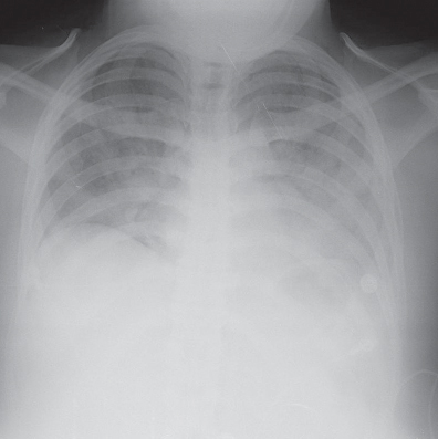
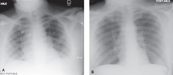
CASE 134 40-year-old woman found down and unresponsive following a heroin drug overdose AP chest radiograph (Fig. 134.1) demonstrates an upper limit normal heart size for the depth of inspiration, bilateral perihilar ground glass opacities, and diminished vascular clarity with poorly defined pulmonary vessels, and dependent layering left pleural effusion. Heroin-Induced Pulmonary Edema • Increased Hydrostatic Pressure Pulmonary Edema of Other Etiologies • Permeability Edema with Diffuse Alveolar Damage (DAD) • Permeability Edema without Diffuse Alveolar Damage (DAD) • Aspiration Pulmonary edema directly associated with an overdose of opiates occurs almost exclusively with heroin but may also occur with the use of cocaine and “crack” (Fig. 134.2). Heroin-induced pulmonary edema complicates 15% of heroin overdose cases. Both cardiogenic and non-cardiogenic pulmonary edema have been reported with intravenous cocaine abuse and crack cocaine smoking. Pulmonary edema is seen in 77–85% of cocaine-related deaths at autopsy. Fig. 134.1 Fig. 134.2
 Clinical Presentation
 Radiologic Findings
 Diagnosis
 Differential Diagnosis
 Discussion
Background
Related posts:
![]()
Stay updated, free articles. Join our Telegram channel

Full access? Get Clinical Tree


134 Heroin-Induced Pulmonary Edema


