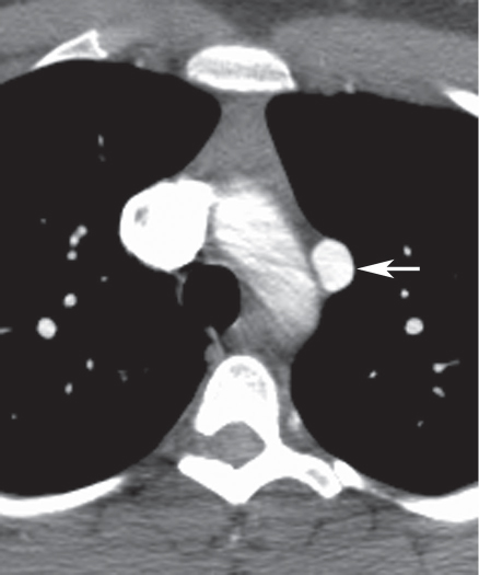
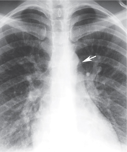
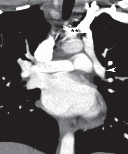
CASE 10 36-year-old woman with cough and fever Coned-down PA chest radiograph (Fig. 10.1) demonstrates an abnormal contour of the left superior mediastinum (arrow). Coronal (Fig. 10.2) and axial (Figs. 10.3, 10.4) contrast-enhanced chest CT (mediastinal window) demonstrates the anomalous course of the left superior pulmonary vein (arrowhead) (Fig. 10.2), which anastomoses with a vertical left superior mediastinal vein (arrow) (Fig. 10.3), which drains into the left brachiocephalic vein (double asterisk). Note the absence of the left superior pulmonary vein from its normal anatomic position posterior to the left atrial appendage (asterisk) (Fig. 10.4) and dilatation of the superior vena cava (Fig. 10.2). Partial Anomalous Pulmonary Venous Return; Left Upper Lobe • Persistent Left Superior Vena Cava Fig. 10.1 (Image 10.1 courtesy of Maysiang Lesar, MD, National Naval Medical Center, Bethesda, Maryland.) Fig. 10.2
 Clinical Presentation
 Radiologic Findings
 Diagnosis
 Differential Diagnosis
10 Partial Anomalous Pulmonary Venous Return