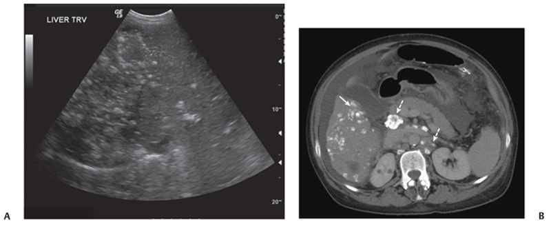
CASE 10 A 56-year-old woman presents with abdominal pain. Fig. 10.1 (A) Ultrasound image shows multiple hyperechoic foci in the liver. (B) Axial non-contrast-enhanced CT demonstrates multiple calcified lesions within the liver (arrow). In addition, calcified lymph nodes are seen in the peripancreatic and retroperitoneal location (dashed arrows). An ultrasound image shows multiple hyperechoic foci in the liver (Fig. 10.1). Axial non -contrast-enhanced computed tomography (CT) demonstrates multiple calcified lesions within the liver. In addition, calcified lymph nodes are seen in the peripancreatic and retroperational location. Calcified hepatic metastases in a patient with breast cancer
Clinical Presentation
Radiologic Findings
Diagnosis
Differential Diagnosis
Discussion
Background
Related posts:


Stay updated, free articles. Join our Telegram channel

Full access? Get Clinical Tree


