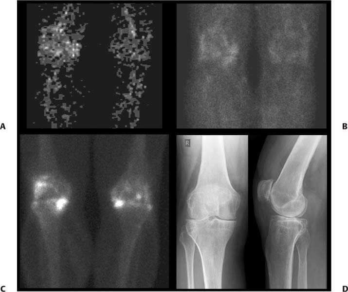
CASE 103 A 56-year-old man with rheumatoid arthritis presents with right knee pain refractory to conventional therapy. Fig. 103.1 • 99mTc-MDP, 25 mCi (925 MBq) intravenously, followed by 60-second dynamic flow (Fig. 103.1A), 60-second pool (Fig. 103.1B), and bone phase imaging 3 hours later (Fig. 103.1C). Flow (Fig. 103.1A) and pool (Fig. 103.1B) images from a 99mTc-MDP bone scan demonstrate significantly increased blood flow to the right knee due to inflammation. The delayed bone phase image from the bone scan (Fig. 103.1C) demonstrates increased uptake, particularly in the medial compartment and the patellofemoral joint. There is increased uptake to a lesser extent in the left knee. Anterior and lateral radiographs (Fig. 103.1D) reveal marked joint space narrowing with minimal osseous proliferation as well as a joint effusion, consistent with the rheumatoid arthritis. Following local anesthesia and sterile preparation, a 22-gauge needle was used to enter the knee compartment from a patellofemoral approach. Straw-colored fluid was aspirated. One milliliter of radio-graphic contrast was injected, and fluoroscopy was performed to confirm the intra-articular position. 90Y, 5 mCi, was then injected into the joint, along with 1 mL of 0.5% bupivacaine and 80 mg of depomethylprednisolone.
Clinical Presentation
Technique
Image Interpretation
Therapy
Discussion
Stay updated, free articles. Join our Telegram channel

Full access? Get Clinical Tree


