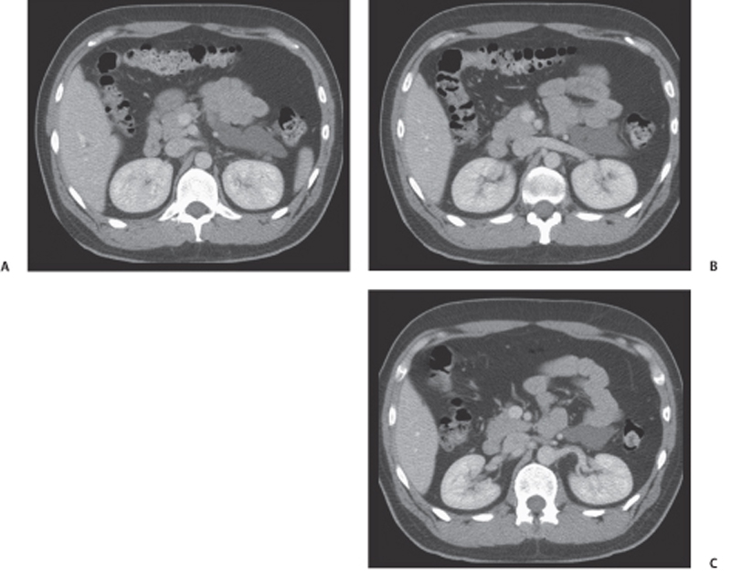
CASE 106 A 34-year-old man is evaluated for chest pain. Fig. 106.1 (A–C) Axial CT images show a lobulated, low-attenuation mass, inferior to the body and tail of the pancreas without enhancement. Axial computed tomography (CT) images reveal a lobulated, low-attenuation mass, inferior to the body and tail of the pancreas without enhancement (Fig. 106.1). Retroperitoneal lymphangioma
Clinical Presentation
Radiologic Findings
Diagnosis
Differential Diagnosis
Discussion
Background
Related posts:


Stay updated, free articles. Join our Telegram channel

Full access? Get Clinical Tree


