CASE 108 Prenatal ultrasonography demonstrated hydronephrosis and oligohydramnios. Immediate postnatal ultrasonography confirms the hydronephrosis and shows posterior urethral valves and a thickened bladder wall. Figure 108A Figure 108B Ultrasonography of the abdomen and pelvis demonstrates a markedly thickened bladder wall (Fig. 108A1), hydroureter, hydronephrosis (Fig. 108A2), and distended posterior urethra. Voiding cystourethrogram shows posterior urethral valves with thickened bladder and diverticulae (Fig. 108B). Posterior urethral valves with thickened bladder wall, hydroureter, and hydronephrosis. A percutaneous nephrostomy was requested. Percutaneous nephrostomy is a well-established procedure for the treatment of obstructive uropathy in the pediatric population. The neonatal population presents some additional difficulties, which are not seen in older children. Placing catheters in neonates may therefore be challenging even when the collecting systems are severely dilated. Congenital malformations are the predominant cause of urinary tract obstruction in the pediatric population. These include:
Clinical Presentation
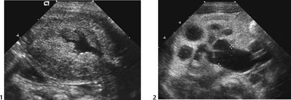
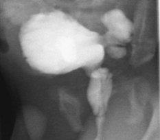
Radiologic Findings
Diagnosis
Differential Diagnosis
Discussion
Background
Etiology
Indications
Related posts:


Stay updated, free articles. Join our Telegram channel

Full access? Get Clinical Tree


