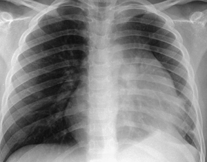
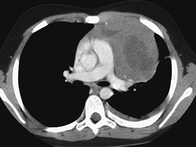
CASE 109 A 9-year-old male presents to his family physician with a cough lasting a few days, shortness of breath with minimal exercise (worse when lying down), lethargy, chest pain, and intermittent fever. Patient is given antibiotics for possible pneumonia and is sent home. The child returns with persisting symptoms. A chest x-ray is ordered, which reveals a mediastinal mass. Figure 109A Figure 109B Chest radiograph demonstrates a left-sided mediastinal mass overlying the heart border (Fig. 109A). Chest CT shows a mass in the anterior mediastinum adjacent to the left cardiac border. The mass is of soft tissue density with central areas of hypodensity (Fig. 109B). Mediastinal mass requiring biopsy subsequently confirmed lymphoblastic lymphoma. The diagnosis of mediastinal masses in children may present a challenge. They are a heterogeneous group and may be of congenital, infectious, or neoplastic origin: A percutaneous core biopsy is a valuable tool in the diagnosis and pathologic characterization of a mediastinal mass. It is especially indicated if imaging suggests that the mass is invasive or unresectable. Four approaches have been used traditionally to biopsy mediastinal masses: parasternal, suprasternal, transpulmonary, and paraspinal. A transsternal approach also can be used in certain cases to biopsy mediastinal masses. Thymoma, neurogenic tumors, and benign cysts represent ~60% of mediastinal masses. Some differences exist between the adult and the pediatric populations. In adults, the most frequent lesions include primary thymic neoplasms, thyroid masses, and lymphomas. In the pediatric population, however, neurogenic tumors, germ cell tumors, and foregut cysts represent ~80% of all cases.
Clinical Presentation


Radiologic Findings
Diagnosis
Differential Diagnosis
Discussion
Background
Etiology
Clinical Findings
Stay updated, free articles. Join our Telegram channel

Full access? Get Clinical Tree


