CASE 111 An 84-year-old woman presents with diarrhea and leukocytosis. Fig. 111.1 (A–D) Axial and coronal images from a contrast-enhanced CT show an enlarged appendix filled with soft tissue density material and concentric calcifications. There is no significant soft tissue stranding in the surrounding mesentery. Additionally, there are coarse, rounded calcifications within the lymph nodes adjacent to the appendix. Axial and coronal images from a contrast-enhanced computed tomography (CT) scan show an enlarged appendix filled with soft tissue density material and concentric calcifications. There is no significant soft tissue stranding in the surrounding mesentery. Additionally, there are coarse, rounded calcifications within the lymph nodes adjacent to the appendix (Fig. 111.1). Porcelain appendix secondary to an appendiceal mucocele
Clinical Presentation
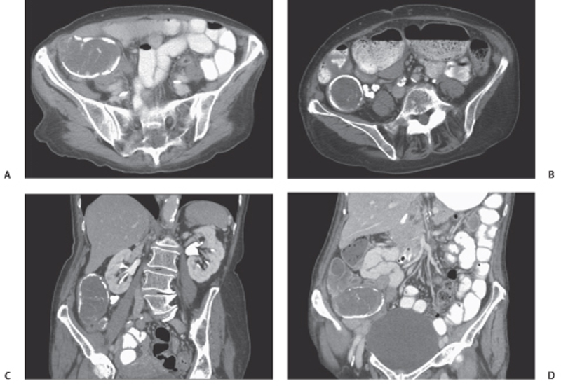
Radiologic Findings
Diagnosis
Differential Diagnosis
Discussion
Related posts:


Stay updated, free articles. Join our Telegram channel

Full access? Get Clinical Tree


