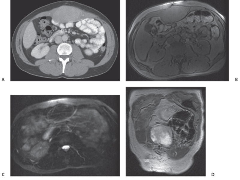
CASE 117 A 30-year-old woman complains of abdominal swelling. Fig. 117.1 (A) Contrast-enhanced axial image shows a moderately enhancing homogeneous lesion in the muscular compartment of the anterior abdominal wall. (B) Noncontrast axial T1-weighted image shows a well-defined midline anterior abdominal wall lesion isointense to muscles in the muscular compartment. (C) Axial T2-weighted image shows a well-defined midline anterior abdominal wall lesion isointense to muscles. (D) Contrast-enhanced coronal T1-weighted image shows an avidly enhancing, well-defined, midline anterior abdominal wall lesion. Postcontrast computed tomography (CT) scan and pre- and postcontrast magnetic resonance imaging (MRI) show a well-defined homogeneously enhancing lesion in the anterior abdominal wall musculature in the midline that is isodense and isointense to muscles (Fig. 117.1). Anterior abdominal wall desmoid tumor (aggressive fibromatosis)
Clinical Presentation
Radiologic Findings
Diagnosis
Differential Diagnosis
Discussion
Background
Related posts:


Stay updated, free articles. Join our Telegram channel

Full access? Get Clinical Tree


