CASE 120 A 45-year-old man presents with right upper quadrant pain (Fig. 120.1). Fig. 120.1 • The patient should take nothing by mouth between less than 24 hours and more than 4 hours before the test. • Immediately following the injection of 1.5 mCi of 99mTc-DISIDA, the patient is placed in a supine position beneath the gamma camera. • Continuous images acquired in a 256 × 256 matrix are divided into 1-minute frames. • Straight anterior views are obtained with a high-resolution, parallel-hole collimator. • Delayed static images may be acquired as necessary. Selected dynamic images show rapid uptake of tracer into the liver and subsequent excretion into the biliary tract and small bowel (Fig. 120.1). The gallbladder is seen within 10 minutes. • Normal study • False-negative study secondary to tracer in duodenum overlying gallbladder fossa (although this would be expected to vary over time) • Bile leak into gallbladder fossa • Acalculous cholecystitis • Choledochal cyst overlying gallbladder fossa Peptic ulcer disease was diagnosed. No evidence of biliary disease was noted.
Clinical Presentation
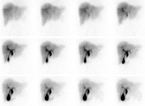
Technique
Image Interpretation
Differential Diagnosis
Diagnosis and Clinical Follow-Up
Discussion
Related posts:


Stay updated, free articles. Join our Telegram channel

Full access? Get Clinical Tree


