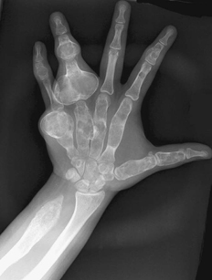
CASE 122 A 14-year-old boy presents with marked enlargement of the left fourth proximal and middle phalanges, left fifth middle phalanx, and ulnar aspect of the left hand. Figure 122A Hand radiograph (Fig. 122A) shows dramatic intramedullary expansion of the left fourth proximal and middle phalanges, as well as the distal aspect of the left fifth metacarpal. Similar, but less dramatic, foci of intramedullary expansion, with endosteal scalloping, involve the left first through fourth metacarpals, the left second and fifth middle phalanges, and the left fourth distal phalanx. In addition, the distal ulna is tapered and hypoplastic, with resultant ulnarization of the carpus (pseudo-Madelung deformity). Affected bones demonstrate an internal “ground-glass” matrix with endosteal scalloping and occasional punctate calcifications, without evidence of an extra-osseous soft tissue mass or sclerotic rim. Ollier disease (multiple enchondromas) Many conditions are associated with focal or diffuse osseous bowing and intraosseous expansile lesions. Patients with Ollier disease, or multiple enchondromas, first described in 1899, present with painless enlargement and deformity of short bones and long bones during childhood. This process is most commonly unilateral; when bilateral, it is usually asymmetrical. Affected long bones are often shortened, whereas affected short tubular bones may be grotesquely expanded and occasionally lengthened. This process is limited to the metaphyses and diaphyses. This is a rare and sporadic entity. As is now recognized, ~20 to 50% of patients will develop malignant dedifferentiation of an intra-osseous lesion over the course of their lifetime; resultant tumors are usually chondrosarcomas. Astrocytomas of the central nervous system may develop in some patients. Patients with Maffucci syndrome demonstrate numerous enchondromas as well as cutaneous and subcutaneous hemangiomas. These patients are also at high risk for osseous and CNS malignancies. Patients with neurofibromatosis may have focal bony expansion, but more commonly they demonstrate narrowing, elongation, and pseudoarthroses of affected long bones. Patients with multiple hereditary exostoses have exophytic expansile lesions; involvement of the distal radius and ulna may also characteristically produce the pseudo-Madelung deformity (Fig. 122B). Some patients demonstrate a mutation in PPR, the receptor for parathormone/parathormone-related peptide. This peptide is involved in maintaining chondrocytes in a nonhypertrophic proliferative state. The macroscopic features of Ollier syndrome include islands of benign resting cartilage within metaphyses and diaphyses, which subsequently proliferate. Microscopically, calcifications, ranging from punctate to “popcorn” in appearance may develop within the cartilaginous matrix. Patients with Ollier syndrome commonly present with painless swelling of hands, wrists, and knees. Wrist and digital motion is often limited, and the long bones may be shortened. This process is usually unilateral; however, when bilateral it is often asymmetric but may also present in a bilateral, symmetric fashion. The pain may result from pathologic fracture or malignant dedifferentiation (usually later in life).
Clinical Presentation
Radiologic Findings
Diagnosis
Differential Diagnosis
Discussion
Background
Etiology
Pathology/Histology
Clinical Findings
Related posts:


Stay updated, free articles. Join our Telegram channel

Full access? Get Clinical Tree


