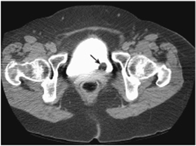
CASE 124 A 46-year-old woman presents with hematuria. Fig. 124.1 Delayed contrast-enhanced axial CT image through the bladder demonstrates a smooth, fat-containing polypoid mass (arrow) arising from the posterior bladder wall. Delayed contrast-enhanced axial computed tomography (CT) image through the bladder demonstrates a smooth, fat-containing polypoid mass arising from the posterior bladder wall (Fig. 124.1). Lipoma of the urinary bladder On cystography and intravenous (IV) urography:
Clinical Presentation
Radiologic Findings
Diagnosis
Differential Diagnosis
Related posts:


Stay updated, free articles. Join our Telegram channel

Full access? Get Clinical Tree


