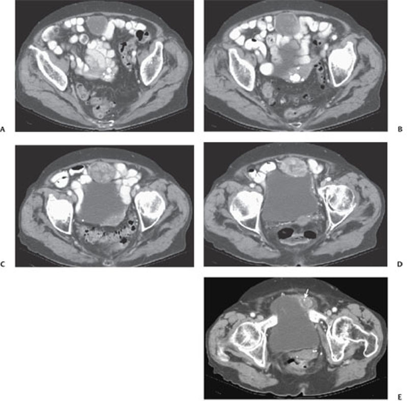
CASE 126 A 47-year-old asymptomatic woman Fig. 126.1 (A–E) Sequential axial contrast-enhanced CT images through the pelvis show a midline, well -defined soft tissue mass overlying the anterosuperior aspect of the bladder containing low-density areas and scattered punctate calcifications (arrow). Axial contrast-enhanced computed tomography (CT) images through the pelvis demonstrate a mid-line, well- defined soft tissue mass overlying the anterosuperior aspect of the bladder containing low-density areas and scattered punctate calcifications (Fig. 126.1). Urachal adenocarcinoma
Clinical Presentation
Radiologic Findings
Diagnosis
Differential Diagnosis
Discussion
Background
Related posts:


Stay updated, free articles. Join our Telegram channel

Full access? Get Clinical Tree


