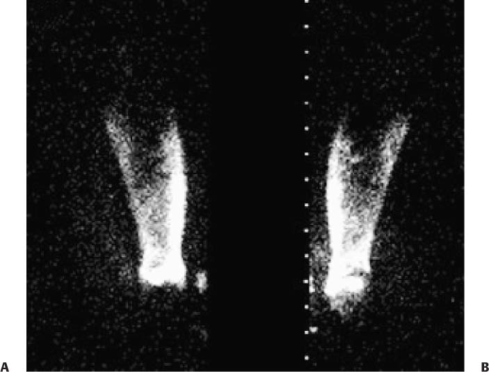
CASE 128 A 50-year-old woman has a history of breast cancer treated with left breast lumpectomy, axillary lymph node dissection, and radiation. She presents with progressive swelling of the ipsilateral arm and forearm. Results of Doppler studies are normal. She is referred for lymphoscintigraphy to evaluate lymphatic drainage. Fig. 128.1 • The radiopharmaceutical is filtered (0.22 μm filter) sulfur colloid labeled with 99mTc-sulfur colloid. It is administered at a concentration of 10 mCi (370 MBq)/mL and at a dose of 0.5 mCi (18.5 MBq) per injection site. • Administer two 0.05-mL intradermal injections of filtered 99mTc-sulfur colloid in the dorsum (as opposed to the web space) of the hand with sterile technique. The contralateral extremity is sometimes injected to provide a comparison. The patient is then asked to exercise the hand and arm to aid lymphatic drainage from the injection site. • Static images of the forearm, arm, and shoulder are acquired for 15 minutes each, with the injection sites left just outside the field of view. Images are obtained over the first 90 minutes after injection or until the lymph vessels and nodal groups are visualized. Delayed views (2–3 hours after the injection) are also obtained to assess for dermal backflow, or “cutaneous flare.” Anterior (Fig. 128.1A) and posterior (Fig. 128.1B) delayed views of the left arm are shown. The hand is outside the lower end of the field of view. The images demonstrate nonvisualization of the deep lymphatic vessels and diffuse dermal backflow, or cutaneous flare, extending from the wrist up to the elbow region. Two faint brachial nodes are seen on the anterior view in the distal aspect of the arm. • Lymphedema • Cellulitis • Edema secondary to a vascular cause
Clinical Presentation
Technique
Image Interpretation
Differential Diagnosis
Diagnosis and Clinical Follow-Up
Stay updated, free articles. Join our Telegram channel

Full access? Get Clinical Tree


