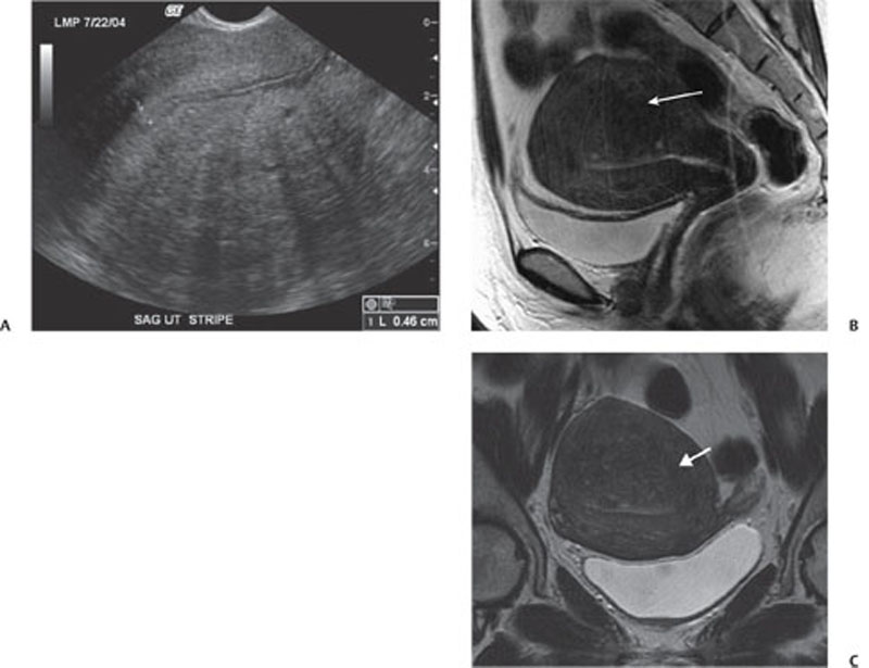
CASE 129 A 53-year-old woman presents with pelvic pain. Fig. 129.1 (A) Transvaginal ultrasound image shows an enlarged uterus with heterogeneous echotexture of the myometrium. (B) Sagittal T2-weighted image of the pelvis shows focal thickening of the junctional zone (arrow). There are T2 hyperintense foci seen in the thickened junctional zone. (C) Coronal T2-weighted image of the pelvis shows a similar finding as the sagittal image (arrow). Transvaginal ultrasound image shows an enlarged uterus with heterogeneous echotexture of the myometrium. Axial and sagittal magnetic resonance (MR) T2-weighted images show a diffusely enlarged uterus with thickening of the junctional zone; small, scattered T2 hyperintense foci are seen within the myometrium (Fig. 129.1A). Adenomyosis Adenomyosis is a common gynecologic disorder, typically occurring in premenopausal multiparous women and characterized at histology by the presence of heterotopic endometrial glands and stroma within the myometrium with compensatory smooth muscle hyperplasia. The ectopic endometrial tissue usually consists of basalis-type cells that are scarcely responsive to hormonal stimuli. Patients usually present with pelvic pain, dysmenorrhea, and abnormal uterine bleeding, although these symptoms are nonspecific and may be seen in other gynecologic disorders, such as leiomyoma, uterine malignancies, and dysfunctional uterine bleeding. In some cases, adenomyosis is associated with infertility. Adenocarcinomas may arise from adenomyosis, but this finding is extremely rare. The etiology is under debate but still unknown. Recent studies demonstrate the central role of tamoxifen, a nonsteroidal antiestrogen agent routinely administered for the treatment of estrogen-sensitive breast cancer.
Clinical Presentation

Radiologic Findings
Diagnosis
Differential Diagnosis
Discussion
Background
Clinical Findings
Complications
Etiology
Stay updated, free articles. Join our Telegram channel

Full access? Get Clinical Tree


