CASE 13 A 28-year-old woman presents with ill-defined abdominal pain. Fig. 13.1 Ultrasound image of the gallbladder showing a well-defined, rounded, hyperechoic nonshadowing polyp in the gallbladder lumen. Ultrasond image of the gallbladder shows a well-defined hyperechoic nonshadowing polyp in the gallbladder lumen (Fig. 13.1). Gallbladder polyp Gallbladder polyps can be benign or malignant. The most common type of gallbladder polyp is a cholesterol polyp, which is a benign condition. Other benign causes of polyps include inflammatory polyps, leiomyoma, fibroma, hemangioma, hamartoma, lipoma, and granular cell neoplasm. Adenoma of gallbladder, which is a premalignant condition, may also present as a polyp. The risk of malignancy correlates with the size of the adenoma, with more chances of lesions with size > 1.2 to 1.5 cm being malignant. Malignant polyps seen in the gallbladder include adenocarcinoma, squamous cell carcinoma, anaplastic carcinoma, and angiosarcoma.
Clinical Presentation
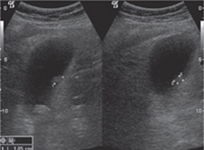
Radiologic Findings
Diagnosis
Differential Diagnosis
Discussion
Background
Related posts:


Stay updated, free articles. Join our Telegram channel

Full access? Get Clinical Tree


