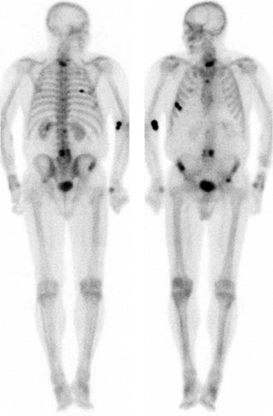
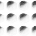
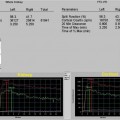
CASE 13 A 64-year-old man with a history of prostate cancer diagnosed 2 years ago now has a prostate- specific antigen (PSA) level elevated to 25 ng/mL (Fig. 13.1). Fig. 13.1 • A 20 mCi dose of 99mTc-MDP is administered intravenously. • Whole-body images of the skeleton are obtained 3 hours after tracer administration. • A 1024 × 256 matrix is used for whole-body images. • Emphasize the importance of oral hydration to improve soft tissue and bladder clearance. Focally intense tracer uptake is seen at several sites, both articular and nonarticular. The injection site is seen at the right antecubital fossa. • Metastatic disease • Trauma Radiographs showed blastic lesions, consistent with prostate cancer.
Clinical Presentation
Technique
Image Interpretation
Differential Diagnosis
Diagnosis and Clinical Follow-Up
Discussion
Stay updated, free articles. Join our Telegram channel

Full access? Get Clinical Tree


