CASE 131
Clinical Presentation
A 36-year-old man presents 18 months after noticing a change in the appearance of a mole on his back. An excisional biopsy performed 3 weeks before this presentation showed a 1.04-mm level 3 malignant melanoma.

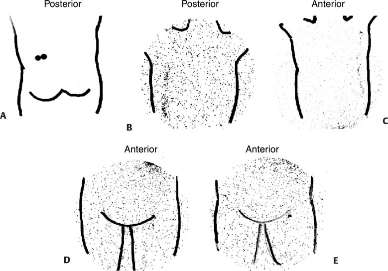
Fig. 131.1
Technique
• Two to four 100- to 200-μCi doses of filtered sulfur colloid (0.22–μm filter) labeled with 99mTc are diluted in a volume of 0.1 to 0.2 mL. The use of 1-mL syringes with 25-gauge needles is recommended. Drawing approximately 0.1 mL of air behind the tracer solution will ensure that the dose is emptied from the syringe during injection.
• For injection, it is helpful to bend the needle to a 45-degree angle using the loosened cap as a sterile tool. The application of standard sterile povidone-iodine (Betadine) followed by an alcohol preparation is recommended.
• Draping the field will reduce the chance of skin contamination.
• Use a high-resolution collimator.
• Energy window 20% centered on 140 keV.
• Imaging time is 60 seconds per view.
• After sterile preparation of the injection site, inject the tracer intradermally at four sites surrounding the lesion and within 1.5 cm of the lesion. Intradermal injections usually raise a “wheal” or bump of tense skin at the injection site. Before withdrawing the needle from the skin, place a 2 × 2 or 4 × 4 gauze pad over the injection site to prevent the injectate from spraying back out of the puncture wound when the needle is withdrawn. Consider SPECT or SPECT/CT for improved localization of the site (s) of tracer uptake.


