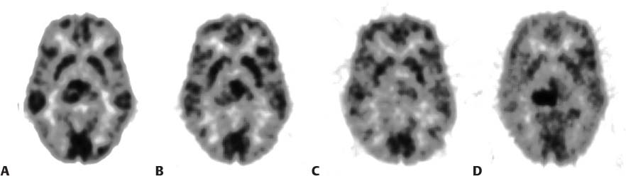
CASE 134 A 25-year-old woman in otherwise good health presents with several episodes of severe headache. MRI demonstrates a small area of enhancement in the right thalamus, and stereotactic biopsy confirms the diagnosis of grade 3 astrocytoma. A 18F-FDG-PET study is performed to define the metabolic extent of the lesion, determine if additional lesions are present, and serve as a baseline for monitoring the effect of radiation therapy. The four images below are axial slices obtained at the same location in the brain at different time points: 134.1A was obtained prior to therapy; 134.1B is mid-way through radiation therapy; 134.1C is done at the completion of therapy; 134.1D is 6 months following the completion of therapy. Fig. 134.1 • The patient should take nothing by mouth for 12 hours before the radiopharmaceutical is administered. Switch all glucose-containing intravenous solutions to normal saline on the day before imaging. • During injection and acquisition, the room should be quiet, with the lights dimmed (avoid disturbances and noise). • 18F-FDG (dose determined by type of imaging system and patient weight) is administered intravenously 45 minutes before image acquisition. • The imaging device is a dedicated PET camera or PET/CT scanner. • Carefully position the patient in a head holder. Before therapy, the transaxial 18F-FDG-PET image of the brain demonstrates an intense focus of increased accumulation in the anterior aspect of the right thalamus (Fig. 134.1A
Clinical Presentation
Technique
Image Interpretation
![]()
Stay updated, free articles. Join our Telegram channel


Full access? Get Clinical Tree


