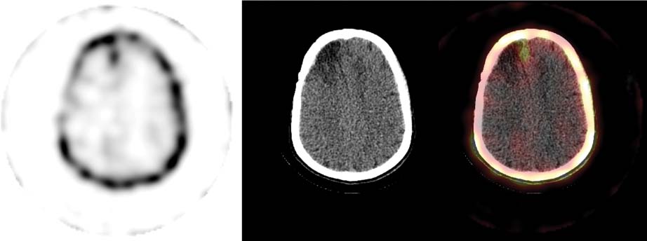
CASE 138 A 26-year-old man in whom a right frontal anaplastic oligodendroglioma was diagnosed 5 years earlier underwent surgical resection initially, followed by radiation therapy. He now presents for repeated imaging with 201TI (Fig. 138.1) because of evidence of recurrence on MRI. Fig. 138.1 • A 3 mCi dose of 201TI is injected intravenously. • Imaging begins 20 to 30 minutes after injection. • Acquisition protocol: 30 minutes with SPECT/CT Images show an abnormal area of increased thallium uptake involving the right frontal region just adjacent to midline (Fig. 138.1). No other areas of abnormally increased thallium accumulation are noted. • Recurrent brain tumor • Cerebral sarcoid (rare) • Meningioma (rare)
Clinical Presentation
Technique
Image Interpretation
Differential Diagnosis
Stay updated, free articles. Join our Telegram channel

Full access? Get Clinical Tree


