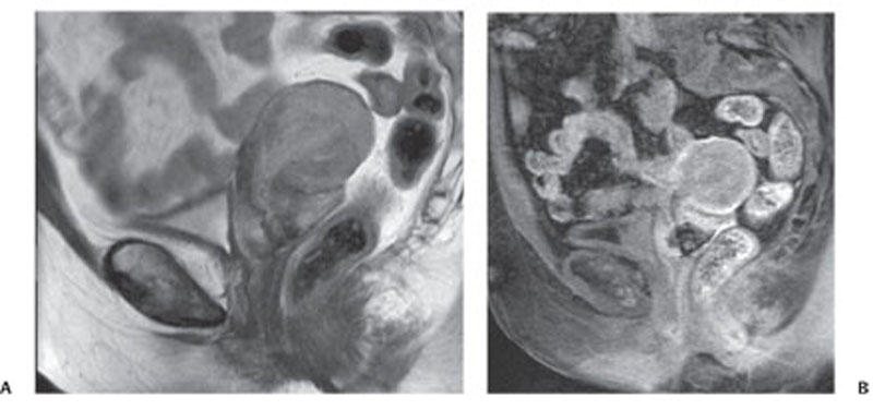
CASE 139 A 65-year-old woman presents with vaginal bleeding. Fig. 139.1 (A) Sagittal T2-weighted image demonstrates a solid mass with low-intermediate signal intensity within the endometrial cavity. The mass extends inferiorly, with infiltration of the cervical stroma, and invades the myometrium superiorly. (B) Sagittal gadolinium-enhanced image shows a relative lack of enhancement of the tumor compared with the myometrium. Sagittal T2-weighted images (Fig. 139.1) demonstrate a solid mass with low-intermediate signal intensity within the endometrial cavity. The mass extends inferiorly, with infiltration of the cervical stroma, and invades the myometrium superiorly. Endometrial adenocarcinoma Endometrial carcinoma is the most common gynecologic malignancy in the United States but only the eighth leading cause of death from cancer in the female population, as it is usually detected at an early stage. This neoplasm mostly affects perimenopausal and postmenopausal women in their 5th to 6th decades, whereas it is uncommonly seen in patients younger than 40 years (< 5%). Nearly 80% of endometrial neoplasms are adenocarcinomas, but other histologic subtypes have been described, including adenosquamous, clear cell, and papillary serous carcinomas. Sarcomas are considerably rarer than adenocarcinomas, but they are associated with a poorer prognosis. Adenocarcinomas may arise from adenomatous hyperplasia of the endometrium (higher risk of progression in cases of cellular atypia); this condition results from the overgrowth of uterine endometrium in postmenopausal women treated with exogenous estrogen or affected by estrogen-producing ovarian neoplasms. Endometrial adenocarcinomas have also been shown to arise from endometrial polyps (~10% are malignant in postmenopausal women); these lesions are usually discovered by accident, although some of them may be associated with vaginal bleeding. The most common symptom observed in up to 90% of patients is postmenopausal vaginal spotting or bleeding. Other presenting symptoms are purulent vaginal discharge, pain, weight loss, and changes in bladder and bowel habits, however, these are symptoms of advanced disease.
Clinical Presentation

Radiologic Findings
Diagnosis
Differential Diagnosis
Discussion
Background
Clinical Findings
Complications
Stay updated, free articles. Join our Telegram channel

Full access? Get Clinical Tree


