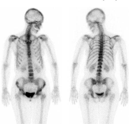
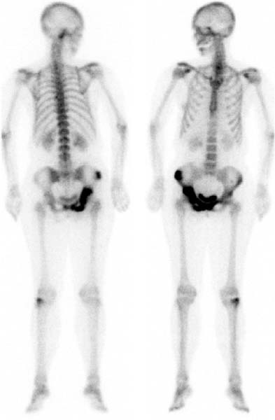
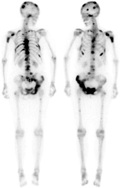
CASE 14 A 43-year-old woman with a history of breast cancer diagnosed 5 years earlier presents with elevated serum levels of calcium and alkaline phosphatase on routine follow-up (Figs. 14.1, 14.2, and 14.3). Fig. 14.1 Fig. 14.2 Fig. 14.3 • A 20 mCi dose of 99mTc-MDP is administered intravenously. • Whole-body images of the skeleton are obtained 3 hours after tracer administration. • A 1024 × 256 matrix is used for whole-body images. • Emphasize the importance of oral hydration to improve soft tissue and bladder clearance. The images show the progression of metastatic disease over approximately 3 years. Figure 14.1, from January 1995, shows right anterior iliac crest involvement and the possibility of pubic symphysis involvement. Figure 14.2, from April 1996, shows definite progression of the disease within the pelvis. Figure 14.3, from April 1997, shows disseminated metastases throughout the axial skeleton, with faint focal abnormalities in the femora as well.
Clinical Presentation
Technique
Image Interpretation
Differential Diagnosis
Related posts:


Stay updated, free articles. Join our Telegram channel

Full access? Get Clinical Tree


