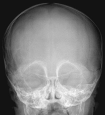
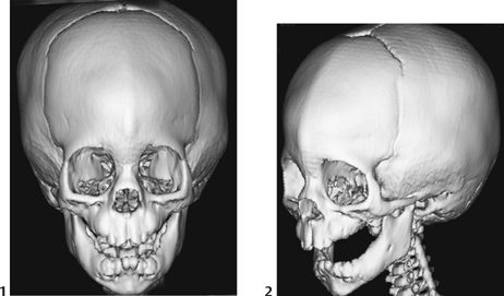
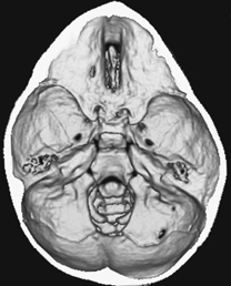
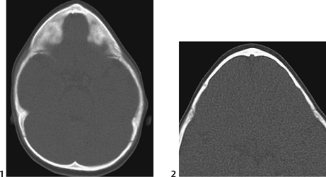
CASE 14 A 7-month-old male presents with triangular appearance of the forehead and flattened temporal fossae, giving a “keel-shaped” appearance to the head. Figure 14A Figure 14B Figure 14C Figure 14D The AP plain x-ray of the skull demonstrates hypotelorism, with medial elevation of the superior orbital rims giving the face a “quizzical” appearance (Fig. 14A). Selected images from 3D CT volumerendered reconstructions: frontal (Fig. 14B1) and oblique (Fig. 14B2) show a prominent, ridged metopic suture. The frontal squama is poorly expanded, in contrast with the temporoparietal widening of the skull. The inside view of the skull (Fig. 14C) demonstrates its triangular shape (trigonocephaly). Note narrowing of the anterior cranial fossa, lateral shortening of the orbital roofs, a deep and narrow cribriform plate groove, and the bilateral bulging and widening of the middle cranial fossa. Axial imaging illustrates the trigonocephaly with the metopic beak (Fig. 14D1) and the endocranial metopic notch in the region of the anterior portion of the superior sagittal sinus (Fig. 14D2).
Clinical Presentation
Radiologic Findings
| Suture Affected | Resulting Deformity | Description |
| Metopic | Trigonocephaly | Triangular skull, keel-shaped, frontal median ridge, symmetrical |
| Coronal, bilateral | Anterior brachycephaly | Skull short anteriorly, wide parietal, symmetrical |
| Coronal, unilateral | Anterior plagiocephaly | Unilateral frontotemporal flattening |
| Sagittal | Scaphocephaly | Elongated, canoe-shaped skull, symmetrical |
| Lambdoid, bilateral | Posterior brachycephaly | Skull short posteriorly, anterior bulge, symmetrical |
| Lambdoid, unilateral | Posterior plagiocephaly | Unilateral posterior flattening |
| All | Oxycephaly | Small rounded skull, symmetrical; needs early surgery to allow the brain to grow |
| All, bone dysplastic |

