86 Renal Vein Thrombosis

CASE 86


Clinical Presentation
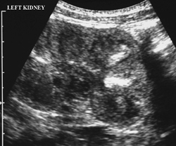
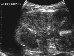

A 3-day-old newborn girl has abdominal ultrasonography for investigation of hematuria, which had been preceded by septicemia.


image

Figure 86A


image

Figure 86B


Radiologic Findings


The left kidney is echogenic on these longitudinal ultrasound images with poor corticomedullary differentiation (Figs. 86A and 86B). Within some of the medullary pyramids, linear areas of increased echogenicity are present, probably representing abnormal interlobar veins. Overall, the left kidney is enlarged. Venous signals from the left kidney were difficult to achieve on Doppler examination. The right kidney is normal.


Diagnosis


Neonatal left renal vein thrombosis (RVT)


Differential Diagnosis


  • Autosomal recessive polycystic kidney disease (causes bilateral enlarged echogenic kidneys with symmetrical appearance)
  • Acute tubular necrosis (usually bilateral and symmetrical without echogenic streaks in the kidneys)
  • Tamm-Horsfall proteinuria (transient bilateral hyperechogenicity of the tips of the medullary pyramids, seen in otherwise well neonates, occasionally precipitated by dehydration)
  • Mesoblastic nephroma (results in a unilateral cystic renal mass, is often exophytic, and is usually associated with some normal preserved renal parenchyma)
  • Multicystic dysplastic kidney (causes unilateral “renal” enlargement, but no normal renal tissue is seen, merely multiple anechoic cysts of varying size)
  • Renal lymphoma (would be a consideration in older children, but is usually bilateral in association with non-Hodgkin’s lymphoma elsewhere)

Discussion


Background

Stay updated, free articles. Join our Telegram channel

Dec 21, 2015 | Posted by in PEDIATRIC IMAGING | Comments Off on 86 Renal Vein Thrombosis

Full access? Get Clinical Tree

Get Clinical Tree app for offline access