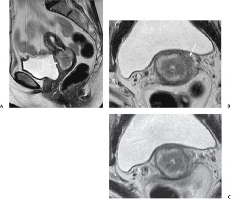
CASE 140 A 56-year-old woman presents with abnormal postmenopausal vaginal bleeding. Fig. 140.1 (A) Sagittal MR T2-weighted image shows a low-intermediate signal intensity mass (arrow) located in the cervix, extending both superiorly to the uterine cavity and inferiorly to the upper third of the vagina. (B,C) Axial MR T2-weighted images demonstrate disruption of the T2 hypointense fibrous stromal ring (arrow) of the parametrium. This finding is suggestive of parametrial invasion (stage IIB). Sagittal and axial magnetic resonance (MR) T2-weighted images (Fig. 140.1) demonstrate a mildly T2 hyperintense mass centered in the cervix. There is evidence of parametrial invasion on the left. Cervical carcinoma Cervical cancer represents the third most common gynecologic malignancy in the United States and the most common gynecologic neoplasm in women age < 45 years worldwide. The incidence has been decreasing in the last few years because of the growing use of screening programs, but it is still high in economically disadvantaged countries. Most cervical neoplasms (90%) are squamous cell carcinomas (SCCs); adenocarcinomas account for the remaining 10% of cases, with different hystopathologic subtypes identified for both of these tumors. SCCs usually spread to the lower uterine segment, vagina, and paracervical spaces along the broad and uterosacral ligament; in advanced stages, progressive involvement of the bladder, rectum, pelvic lymph nodes, and pelvic side wall is common. Radiologic staging plays a central role in patients’ management, as it affects both treatment and outcome. Clinical presentation is correlated strongly with the cervical cancer stage; patients may be asymptomatic (typically in the early stages) or may present with abnormal vaginal bleeding (irregular menses, hypermenorrhea, or painless metrorrhagia) or abnormal vaginal discharge (watery, mucoid, or purulent). Abdominal and pelvic pain, along with rectal or urinary symptoms, usually occurs in advanced stages.
Clinical Presentation

Radiologic Findings
Diagnosis
Differential Diagnosis
Discussion
Background
Clinical Findings
Complications
Stay updated, free articles. Join our Telegram channel

Full access? Get Clinical Tree


