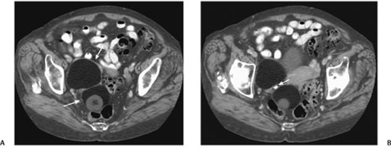
CASE 141 Computed tomography (CT) scans of the chest, abdomen, and pelvis were obtained in a 73-year-old woman as part of a staging work-up for a recently diagnosed lung cancer. Fig. 141.1 Contrast-enhanced CT scans reveal (A) a large right ovarian mass with macroscopic fat (arrows), soft tissue components, and (B) a focus of calcification (arrow). Contrast-enhanced CT reveals a large right ovarian mass with macroscopic fat, soft tissue components, and a focus of calcification (Fig. 141.1). Mature cystic teratoma (dermoid cyst) Ovarian teratomas are the most common germ cell neoplasm and are subdivided into mature cystic teratomas (dermoid cysts), immature teratomas, and monodermal teratomas (i.e., struma ovarii and carcinoid tumors). Mature cystic teratomas are the most prevalent subtype.
Clinical Presentation
Radiologic Findings
Diagnosis
Differential Diagnosis
Discussion
Background
Clinical Findings
Related posts:


Stay updated, free articles. Join our Telegram channel

Full access? Get Clinical Tree


