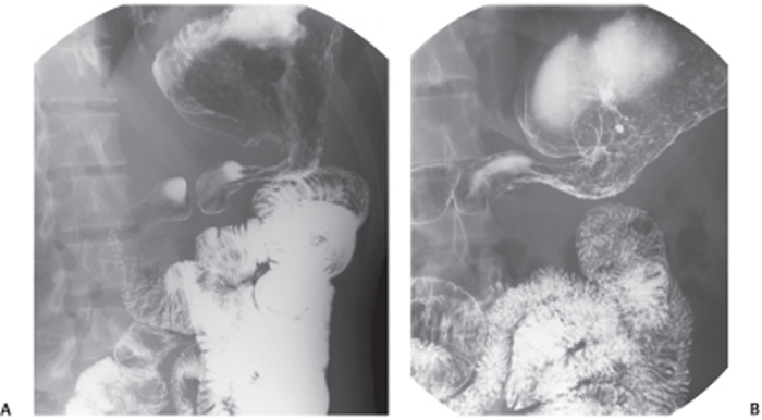
CASE 79 A 28-year-old man complains of epigastric pain, anorexia, and weight loss. Fig. 79.1 (A,B) Axial contrast-enhanced CT images of the abdomen show mild enhancement and diffuse increased thickness of the gastric walls; these findings are suspicious for gastric neoplasm, although an inflammatory process cannot be excluded. Multiple hypodense cystic lesions are seen in the liver and both kidneys. Axial contrast-enhanced computed tomography (CT) images of the abdomen (Fig. 79.1) demonstrate mild and diffuse thickening of the gastric walls. The images from the upper gastrointestinal (GI) series (Fig. 79.2) demonstrate abnormal and persistent narrowing of the distal stomach. Gastric adenocarcinoma Gastric carcinoma is a deadly disease, with decreasing incidence in the past 50 years in North America and Western Europe owing to the earlier identification at imaging and endoscopy. Currently, the incidence of gastric carcinoma is extremely variable worldwide, with the highest number of cases in Japan, China, Russia, and Eastern Europe (> 30 cases per 100,000); men are usually 2 times more commonly affected than women, with a peak incidence in those ages of 50 to 70 years. Fig. 79.2 (A,B) Upper gastrointestinal series images (double-contrast barium examination) demonstrate an abnormally narrowed and nondistensible gastric outlet suspicious for gastric malignancy. No discrete ulceration is identified. Different histologic types have been described: adenocarcinomas (intestinal and diffuse types) account for almost 95% of cases, and lymphomas, leiomyosarcomas, carcinoids, and sarcomas for the remaining 5%. Adenocarcinomas have been classified into four different subtypes: papillary, tubular, mucinous, and signet ring. Almost 30% of gastric malignancies arise from the antrum, 30% from the body, and 40% from the fundus and cardia region. Clinical presentation mostly depends on the disease stage; patients may be asymptomatic (early stage) or may present with epigastric pain, anorexia, weight loss, nausea, vomiting, and upper GI bleeding (hematemesis, melena, occult blood in stools).
Clinical Presentation
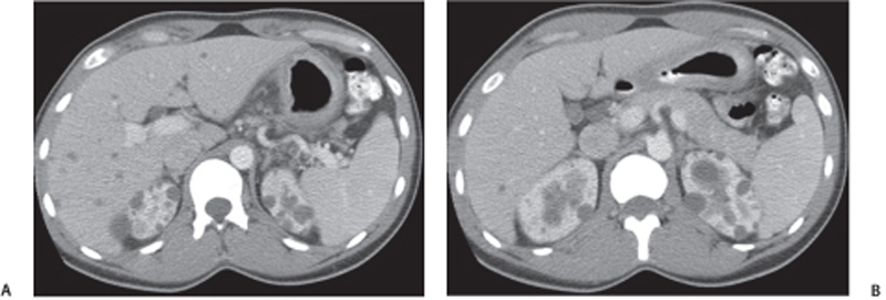
Radiologic Findings
Diagnosis
Differential Diagnosis
Discussion
Background
Clinical Findings
Complications
Related posts:


Stay updated, free articles. Join our Telegram channel

Full access? Get Clinical Tree


