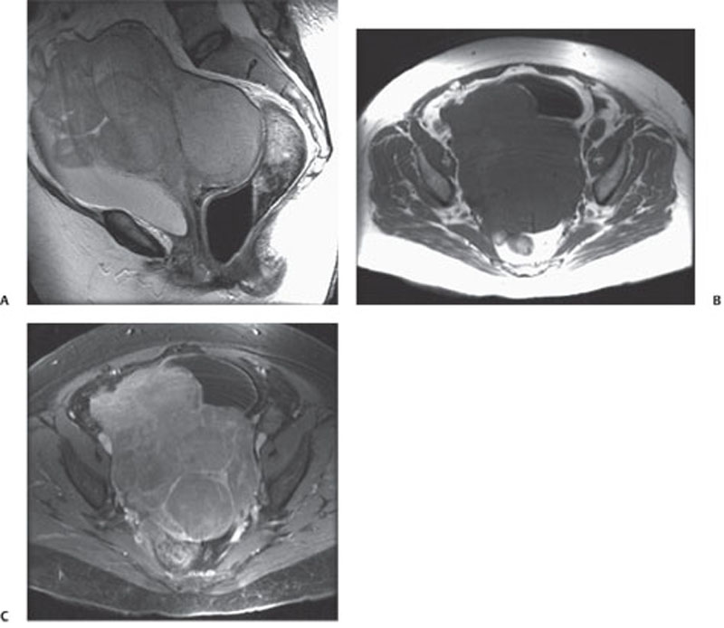
CASE 142 A 67-year-old woman presents with subacute abdominal pain, constipation, and increased abdominal girth. Fig. 142.1 (A) Sagittal T2-weighted image reveals a bulky, lobulated mass centered in the uterine cervix with intermediate signal. (B,C) Transverse T1-weighted images show the mass with homogeneous low signal, which demonstrates moderate, fairly uniform enhancement. A sagittal T2-weighted image (Fig. 142.1A) reveals a bulky, lobulated mass centered in the uterine cervix with intermediate signal. Transverse T1-weighted images (Fig. 142.1B,C) show the mass with homogeneous low signal, which demonstrates moderate, fairly uniform enhancement. Cervical lymphoma (large B-cell, follicular type)
Clinical Presentation

Radiologic Findings
Diagnosis
Differential Diagnosis
Discussion
Background
Stay updated, free articles. Join our Telegram channel

Full access? Get Clinical Tree


