CASE 15 An 8-month-old baby presents with anorectal malformation and sensorimotor deficit of the lower extremities. Figure 15A The sagittal T1-weighted (Fig. 15A1) and T2-weighted (Fig. 15A2) MRI demonstrates agenesis of the lower sacrum and coccyx. The caudal spinal cord is dysplastic and wedge-shaped (arrow). This is due to the agenesis of the conus, which is metamerically consistent with the missing spine. There also is hydrosyringomyelia, and a lipoma of the filum terminale is noted (arrow) (Figs. 15A1 and 15A3). The axial T1-weighted MRI (Fig. 15A3) demonstrates a horseshoe kidney. Partial sacral agenesis, which is part of the so-called caudal regression syndrome Caudal regression syndrome is a rare congenital defect, characterized by the absence of the caudal-most vertebrae and spinal cord. The defect may affect the caudal sacrum only, or it may extend upward as far as the thoracic spine, with functional defects and associated malformations in proportion with the extent of the agenesis. It may be associated with a fusion, and posterior rotation, of the lower limbs (“mermaid” appearance or sirenomelia). Other visceral malformations, especially of the pelvic organs, are common. The pathogenesis of the disorder is poorly understood. Maternal diabetes is a common factor, and insulin in excess has been shown to generate a similar malformation in the chick embryo. The role of segmentation gene defects is likely as well, as it is in other segmental spinal dysgeneses. Embryology holds that the cord and spine develop from two different, successive steps. The first is primary neurulation
Clinical Presentation
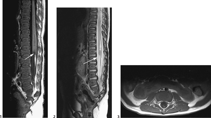
Radiologic Findings
Diagnosis
Differential Diagnosis
Discussion
Background
Embryology
![]()
Stay updated, free articles. Join our Telegram channel


Full access? Get Clinical Tree


