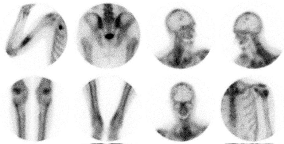
CASE 15 A 65-year-old man with non–small-cell lung carcinoma undergoes bone scan for staging (Fig. 15.1). Fig. 15.1 • A 20 mCi dose of 99mTc-MDP is administered intravenously. • Whole-body images of the skeleton are obtained 3 hours after tracer administration. • A 1024 × 256 matrix is used for whole-body images. • A 256 × 256 matrix is used for spot views. • Emphasize the importance of oral hydration to improve soft tissue and bladder clearance. Selected spot views show focally increased activity in the right distal humerus, the left proximal humerus, the skull, and a right upper rib anteriorly. Also noted is diffusely increased activity in the femora and tibiae bilaterally in a “tram track” cortical pattern, consistent with hypertrophic osteoarthropathy (HO). • For the focal bony findings
Clinical Presentation
Technique
Image Interpretation
Differential Diagnosis
 Metastatic disease
 Trauma
Stay updated, free articles. Join our Telegram channel

Full access? Get Clinical Tree


