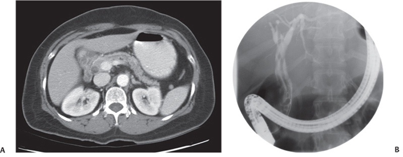
CASE 16 A young man complains of right upper quadrant pain and jaundice. Fig. 16.1 (A) Axial contrast-enhanced CT image shows a dilated common bile duct with a filling defect. (B) Endoscopic retrograde cholangiopancreatography performed subsequently shows multiple linear filling defects with dilatation of the common bile duct. An axial contrast-enhanced computed tomography (CT) image shows a dilated common bile duct with a filling defect. Endoscopic retrograde cholangiopancreatography (ERCP) performed subsequently shows multiple linear filling defects with dilatation of the common bile duct (Fig. 16.1). Biliary ascariasis Infestation with Ascaris lumbricoides (roundworm) is one of the most common helminthic infestations and is highly endemic in developing tropical and subtropical countries. Clinical disease is mostly restricted to subjects with heavy worm loads. The manifestations of ascariasis vary and include constitutional symptoms, particularly pulmonary and gastrointestinal complaints. Hepatobiliary and pancreatic ascariasis can cause distinct clinical presentations: biliary colic, acalculous cholecystitis, acute cholangitis, acute pancreatitis, and hepatic abscess.
Clinical Presentation
Radiologic Findings
Diagnosis
Differential Diagnosis
Discussion
Background
Clinical Findings
Complications
Etiology
Related posts:


Stay updated, free articles. Join our Telegram channel

Full access? Get Clinical Tree


