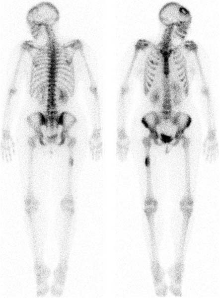
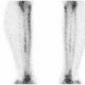
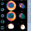
CASE 16 A 31-year-old woman presents with back pain. The patient was referred to a neurologist after a chiropractor noted a spine lesion on plain films. CT and MRI demonstrate a large mass in the left kidney. A bone scan is obtained to demonstrate the extent of metastasis (Fig. 16.1). Fig. 16.1 • A 20 mCi dose of 99mTc-MDP is administered intravenously. • Whole-body images of the skeleton are obtained 3 hours after tracer administration. • A 1024 × 256 matrix is used for whole-body images. • A 256 × 256 matrix is used for spot views. • Emphasize the importance of oral hydration to improve soft tissue and bladder clearance.
Clinical Presentation
Technique
Image Interpretation
Related posts:


Stay updated, free articles. Join our Telegram channel

Full access? Get Clinical Tree


