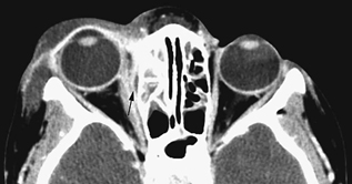
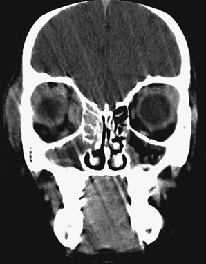
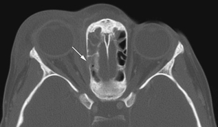
CASE 17 A 4-year-old female presents with right eye swelling and is unable to open her eyelid. She has mild right proptosis. Figure 17A Figure 17B Figure 17C Axial contrast-enhanced CT image through the orbits at the level of the mid globes shows right preseptal soft tissue swelling with preseptal cellulitis and proptosis of the globe (Fig. 17A). This cellulitis also extends laterally along the superficial soft tissues of the temporal region of the face. Extensive ethmoid sinusitis (mucosal enhancement and thickening) is present. There is a small, lentiform subperiosteal fluid collection, with enhancing rim at the medial aspect of the right orbital wall (Fig. 17A, arrow). No intraconal fat stranding is present. On the contrast-enhanced coronal image of the orbits through the midposterior globe, the extent of the subperiosteal fluid collection, with enhancing rim, is better appreciated, as is its relationship to the lamina papyracea and the laterally deviated, slightly thickened medial rectus muscle (Fig. 17B). This fluid collection is adjacent to the lamina papyracea, which has undergone focal disruption (Fig. 17C, arrow). Subperiosteal orbital abscess The clinical diagnosis of orbital and periorbital infections mandates a thorough history, physical examination, and ophthalmologic evaluation. CT scanning assists in the diagnosis of orbital infections and their complications. Demonstration of orbital and/or subperiosteal abscess mandates surgical drainage. Orbital infections have been classified with a widely accepted system proposed by Chandler et al (1970). This five-stage classification system of worsening infections and prognosis is as follows: Stages III and higher are usually treated surgically, with accompanying antimicrobial therapy, usually by empiric intravenous antibiotics, followed by oral antibiotics. Stages I and II are usually treated with antibiotics alone and are followed to ensure resolution.
Clinical Presentation
Radiologic Findings
Diagnosis
Differential Diagnosis
Discussion
Background
Classification
Etiology
Related posts:


Stay updated, free articles. Join our Telegram channel

Full access? Get Clinical Tree


